CRIT CLOUD

  • Summaries & Reviews
    • Airway
    • Cardiovascular
    • Controversies
    • Endocrinology
    • Fluids
    • Guidelines
    • Infections
    • Neurology
    • Nutrition
    • Pharmacology
    • Procedures
    • Radiology
    • Renal
    • Resuscitiation
    • Respiratory
    • Sedation
    • Sepsis
    • Transfusion
  • World of Foam
  • FOAM.education
  • Education
    • ACLS Training ICU Lindenhofspital
    • Download Presentations
    • Download CME Galway Clinic 🔒
    • Download CME Lindenhof Hospital 🔒
    • Echo 🔒
    • Presentations🔒
    • Multimedia
  • About Us
    • Contact

Reviews and Summaries

7 Reasons for the Use Vasopressors through Peripheral Catheters

16/12/2019

 
Picture

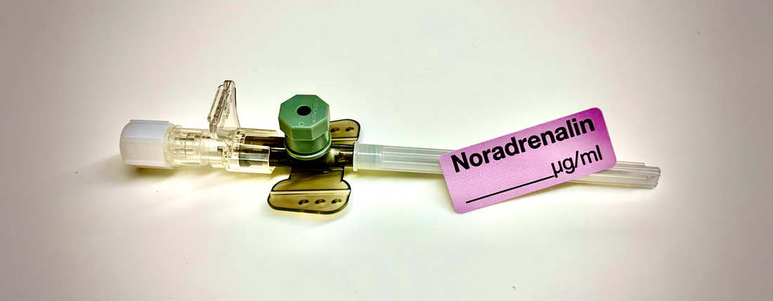
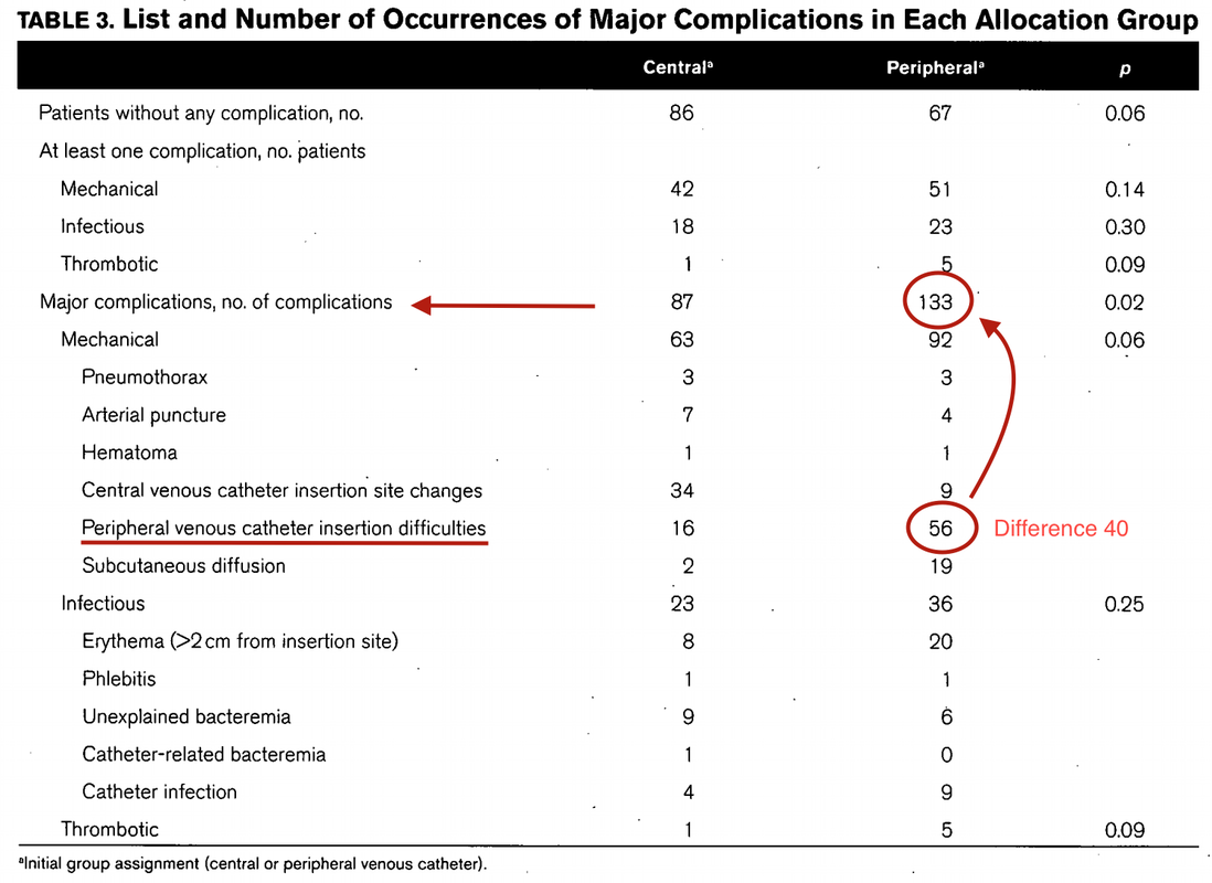
​Teaching in medical school and opinions in literature are in agreement: The application of vasopressors requires central venous access. The reason for this are concerns that vasopressors given over a peripheral venous catheter (PCV) may cause phlebitis or even worse necrosis or ischemia through extravasation. 

​While irritation of a peripheral vein is often observed with the administration of drugs like potassium or amiodarone, this usually is not the case with the application of, e.g. norepinephrine. Besides, it is essential to keep in mind that the insertion of a central venous catheter (CVC) is technically demanding and takes a certain amount of time when performed correctly. The procedure is also associated with potentially dangerous complications that might be hazardous to the patient.

Therefore a fundamental question arises:

Do all patients that require vasopressors need a central venous catheter?
​

​

What about the peripheral access (PVC) - Any dangers there?


​Study #1

In 2015 Cardenas-Garcia et al. have published a 

​open-label, single-centre trial 

in which they treated

a total of 734 patients with the vasopressors noradrenaline (506), dobutamine (101 and phenylephrine 176 via peripheral access only.

The average duration of infusion was 49 hours.

They found


extravasation in only 2% of all patients without any further tissue injury following treatment with local phentolamine injection and nitroglycerin paste.
These findings indicate that:
​
  • Correctly applied vasopressors via a peripheral line are safe, even if given over several hours
  • Complications like extravasation are generally rare and are unlikely to cause any further harm​

J Hosp Med. 2015 Sep;10(9):581-5. doi: 10.1002/jhm.2394. Epub 2015 May 26.


​Study #2

In 2015 Loubani et al. performed a systematic review of extravasation and local tissue injury from the administration of vasopressors through peripheral intravenous catheters and central venous catheters. They looked at
​
  • Local tissue injury close to the infusion site
  • Extravasation of a vasopressor into surrounding tissue or a body cavity
  • Major disability of the patient

An excellent summary of this study can be found on REBELEM, who correctly states that this review was only for complications from administration of vasopressor, and not a review of the frequency of complications (i.e. instances where no complications occurred).

This review shows nicely though that
​
  • Most complications concerned peripheral IV-lines distal to the antecubital or popliteal fossae, and
  • Almost all occurred in infusions running for more than 4 hours

J Crit Care. 2015 Jun;30(3):653.e9-17. doi: 10.1016/j.jcrc.2015.01.014. Epub 2015 Jan 22


​Study #3

In 2017 Lewis at al. performed a retrospective chart review of 202 patients who received vasopressors through a PVL. The primary vasopressors used were norepinephrine and phenylephrine. The most common PVL sites used were the forearm and antecubital fossa. The incidence of extravasation was 4%. All of the events were managed conservatively; none required an antidote or surgical management. Although with many limitations to this review, there is further evidence indicating:

  • Extravasation seems to be a rather rare complication and again did not result in any further harm for the patient

J Intensive Care Med. 2017 Jan 1:885066616686035.


​Study #4

In 2018 Medlej et al. tried to determine the incidence of complications of running vasopressors through PIVs in patients with circulatory shock in a prospective, observational trial. Again, REBELEM has nicely summarized this rather small trial. It is another small indicator that:
​
  • In patients with shock, the use of peripheral vasopressors (noradrenaline and dopamine) in a large bore PVC at a proximal site for less than 4 hours is safe!

​J Emerg Med. 2018 Jan;54(1):47-53.


​Well, how do PVC's compare to CVC's then?


​Study #5

In 2018 Ricard JD et al. performed a

Multicenter, controlled, parallel-group, open-label randomized trial

in which

Patients were randomized to receive central venous catheters (135 patients) or peripheral venous catheters (128 patients) as initial venous access.

The primary endpoint was the rate of major catheter-related complications within 28 days.

They found significantly more PVC-related complications per patient when only treated with peripheral lines compared to patients that received at least one CVC.


And they concluded: "central venous catheters should preferably be inserted: a strategy associated with less major complications"
Picture

REALLY? Hold on! - let's have a close look at those 'major complications, the PRIMARY endpoint of this study!

Picture

Although going through this article several times, it remains difficult to understand how PVC insertion difficulties are comparable major complications.
First of all, difficult venous access is one of the indications for the insertion of a CVC, not its complication. Patients were randomly allocated in a one-to-one ratio to receive a CVC or a PVC. So how can difficult peripheral access be a complication when going for central access directly?
​
Also, there is considerable doubt whether the occurrence of a pneumothorax can be used to compare complications of these two procedures!


However, when eliminating difficult peripheral access as an indication, there is not much left to say PVCs are associated with more complications than CVCs. Moreover, most clinicians will agree that catheter infections in PVCs are less problematic than when occurring in CVCs.

Given these considerations, it seems safe to say:


  • In critically ill patients peripheral access can be tricky indeed
  • PVCs might be associated with more frequent local erythema and extravasation of fluids
  • Good to know: peripheral access is not associated with more pneumothoraces ; )

Crit Care Med. 2013 Sep;41(9):2108-15 


​2019 - More Evidence Keeps Rolling In!
​

Study #6

Tian et al. have performed a 

Systematic review 

in order to assess

 the frequency of complications associated with the delivery of vasopressors via PVCs.

They included

Studies of continuous infusions of vasopressor medications (noradrenaline, adrenaline, metaraminol, phenylephrine, dopamine and vasopressin) delivered via a PiVCs that included at least 20 patients. This resulted in seven observational studies (only) with a total of 1384 patients.

They found that

Extravasation occurred in 3.4% (95% CI 2.5-4.7%) of patients. There were no reported episodes of tissue necrosis or limb ischaemia. All extravasation events were successfully managed conservatively or with vasodilatory medications.


  • Extravasation seems to be an issue with PVCs, but there is no further information on the size or location of the peripheral line.
  • Again, no serious side effects were reported, indicating that peripherally administered vasopressors are safe over all when given for a limited duration.

Emerg Med Australas. 2019 Nov 7.


​Study #7

Pancaro et al. published

a retrospective cohort study

in which  identified


14'385 surgical patients who received peripheral norepinephrine infusions perioperatively with a concentration of 20 µg/mL (a rather low concentration)

They found

Extravasation of norepinephrine in only 5 patients and there where zero related complications requiring surgical or medical intervention. The median time of norepinephrine infusion among these patients was 20 minutes.
This is a fairly good indicator that:

  • Giving vasopressors through PVC for a limited duration is safe
  • Extravasation might actually be harmless when applied in rather lower contentrations

Anesthesia & Analgesia. SEPTEMBER 27, 2019

Picture

Giving the current evidence available, it seems appropriate to conclude:

  • The need for vasopressors itself is not a mandatory indication for central venous access
 
  • Vasopressors can be safely given through a peripheral venous catheter
    • This is especially true when used for a limited time (e.g. less than 4 hours) and when applied in rather lower concentrations.
 
  • ​In the critically ill central venous access will inevitably still be required (advantage of multiple lumens, difficult peripheral access, other drugs that do entitle the use of a  CVC etc.)
​

Has Levosimendan Failed? - A Review of Current Evidence

11/5/2019

 
Picture

​Acute decompensated heart failure comes in many ways as remains a challenge for optimal treatment since various conditions can cause it, e.g. advanced chronic heart failure, cardiogenic- and septic-shock, cardiac- and non-cardiac surgery, etc. Among many other interventions, the infusion of positive inotropes is often used on one side and vasodilators on the other side to stabilize the situation and improve cardiac function. Despite all these measures, the re-admission rate and mortality remain a significant problem among these patients. 

While Levosimendan is still under investigation in the U.S., it is used worldwide for the short-term treatment of acutely decompensated severe chronic heart failure, especially when other treatment options have failed. Some recommend the usage of this drug in a different setting like sepsis-related heart failure or coronary heart disease.


How does Levosimendan work?


Levosimendan has some remarkable properties, which are mainly caused by three mechanisms:

1. Enhancement of the calcium sensitivity of the myofilament by binding to troponin C.

2. Opening of adenosine triphosphate-sensitive potassium (KATP) channels in vasculature smooth muscle.

3. Opening of mitochondrial KATP channels.


These mechanisms result in a positive inotropy of the heart, an increase in stroke volume (SV) and therefore, cardiac output (CO). Besides, it's vasodilatory properties seems beneficial for coronary perfusion and reduce pulmonary capillary wedge pressure (PCWP).

All these effects do not appear to induce an unfavourable energy balance in the myocardial cell, and also the oxygen demand is not increased.

Picture
ScienceDirect.com


​So what's the Evidence?
​
The Small Bits and Pieces


​First publications back in the 90ies and early 2000s were indicative for the properties of levosimendan.  Three studies were randomized and double-blinded but very low in patient numbers and therefore not powered enough to provide substantial evidence. Their statements were also not concerning patient outcome or mortality, but its results confirmed that levosimendan enhances cardiac output without oxygen wasting, is well tolerated and leads to favorable hemodynamic effects.


Lilleberg et al. Eur Heart J. 1998;19:660–668.

Ukkonen et al. Clin Pharmacol Ther. 2000;68:522–531.

Nieminen et al. J Am Coll Cardiol. 2000;36:1903–1912.


In 2003 Kivikko et al. published further data from another small trial indication that its beneficial effects (decreases in left and right heart filling pressures and in SVR, as well as increases in stroke volume and cardiac index) are maintained for at least 24 hours after discontinuation of a 24-hour infusion. At this point, it is worth mentioning that the author published these findings as a current employee of Orion Pharma, which manufactures levosimendan.

Kivikko et al. Circulation. 2003;107:81–86.


Further data indicated that its effect might be sustained for up to at least a week, although also this study included only 22 patients!

8. Lilleberg et al. Eur J Heart Fail. 2007;9:75–82.


A Lancet publication in 2002 by Follath et al. presented a multicentre, randomized, double-blind trial in which they compared the effect of levosimendan to dobutamine in patients that were admitted to a hospital with low-output heart failure and were judged to require hemodynamic monitoring and treatment with an intravenous inotropic agent. In these 203 patients, levosimendan had a consistently better effect than dobutamine on the individual hemodynamic variables at the end of the 24 h treatment period (cardiac output and pulmonary capillary wedge pressure). The change in clinical symptoms like fatigue and dyspnoea were not significantly different, though. 
​

Interestingly, for the first time, the levosimendan group showed lower mortality than in the dobutamine group for up to 180 days.​ However, it's important to mention its absence of placebo control and its rather small sample size.

Follath et al. Lancet. 2002;360:196–202.


The Big Lumps

The SURVIVE Study


In the SURVIVE study Mebazaa et al. compared the efficacy and safety of intravenous levosimendan or dobutamine in patients hospitalized with acute decompensated heart failure who required inotropic support.
They performed a

​randomized, double-blind trial at 75 centers in 9 countries

in which they evaluated

1327 patients hospitalized with acute decompensated heart failure who required inotropic support

They found that

the addition of levosimendan to standard therapy resulted in fewer deaths compared with dobutamine, especially in the first few weeks after treatment

but

Levosimendan did not significantly reduce all-cause mortality at 180 days or affect any secondary clinical outcomes
Mebazaa et al. JAMA. 2007 May 2;297(17):1883-91.


The REVIVE Studies - Heart Failure


In the Randomized Multicenter Evaluation of Intravenous Levosimendan Efficacy studies (REVIVE I and REVIVE II) the efficacy of levosimendan on symptoms of heart failure during five days after starting a 24h trial drug infusion was assessed. 

Each patient's clinical course over 5 days was determined by a composite of the patient's self-assessment of symptoms together with a physician's assessment of the occurrence of clinical deterioration. 
​
The primary endpoint of the study was a new clinical composite endpoint (improvement, unchanged or worse - including death) derived from studies in chronic heart failure and first evaluated in 100 ADHF patients in the REVIVE I pilot study.


In REVIVE II the investigators performed one of the

first large, prospective, randomized, double-blind, controlled trials

in which they evaluated

600 patients  admitted at 103 sites in the United States, Australia, and Israel

with 

worsening heart failure and dyspnea at rest despite treatment with intravenous diuretics, and left ventricular ejection fraction of < 35% measured within the last year

whereas

patients were randomized to either receive levosimendan for 24h or standard treatment alone​

They found that after 5 days

- more patients receiving levosimendan experienced improvement compared with those who were on placebo (19.4% vs 14.6%, respectively, a 33% relative increase; P = .015)

- fewer patients receiving levosimendan worsened compared with patients who were on placebo (19.4% vs 27.2%, respectively), a 29% relative decrease, and

- fewer patients receiving levosimendan required rescue therapy (15.1%) vs placebo (26.2%), a 42% relative decrease.


Among other secondary endpoints, levosimendan improved

B-type natriuretic peptide (BNP) levels, length of hospital stay and dyspnoea at 6 hours

But

 mortality at 90 days did not differ significantly between treatment arms (a secondary endpoint) and

the most common treatment-emergent cardiovascular adverse events were more frequent with levosimendan, including hypotension (50% vs 36%), ventricular tachycardia (25% vs 17%), and atrial fibrillation (8% vs 2%).​

JACC Heart Fail. 2013 Apr;1(2):103-11. doi: 10.1016/


The LeoPARDS-Trial - Sepsis


In this study investigators wanted to know whether in adult patients who have sepsis the application of levosimendan reduces the incidence and severity of acute organ dysfunction compared with placebo.
They performed a

Randomised, double-blind, placebo-controlled multi-centre trial in 34 general ICUs in the UK

in which they evaluated

516 patients with suspected or confirmed infection and 2 or more SIRS criteria who were dependent on vasopressors for at least four hours to maintain their blood pressure.

They compared 

the intravenous infusion of levosimendan or placebo for 24 hours in addition to standard care

and found:

- No significant difference in the daily Sequential Organ Failure Assessment (SOFA) score up to day 28 (primary outcome)


- No statistical difference on death at 28 days, at ICU discharge and hospital discharge (secondary outcome)

- And: The use of levosimendan was associated with more hemodynamic instability, lower mean arterial pressures, therefore more need for noradrenalin at 24h and significantly more supraventricular tachyarrhythmias (all secondary outcomes).
N Engl J Med 2016; 375:1638-1648
​


​The CHEETAH Trial - Cardiac Surgery


The question in CHEETAH was if levosimendan compared to placebo reduces mortality in patients undergoing cardiac surgery with left ventricular dysfunktion.


This time Landoni et al. performed a

Multi-centre, randomised, placebo-controlledand parallel group designed trial 

in which they evaluated 

506 patients scheduled for cardiac surgery with peri-operative cardiovascular dysfunction 

defined as

pre-operative left ventricular ejection fraction (LVEF) < 25%, pre-operative intra-aortic balloon pump (IABP), intra- or post-operative (within 24 hours) IABP or significant inotropic requirement

They compared

Levosimendan infusion continued for up to 48 hours (or until ICU discharge) or placebo

and found

No difference in mortality at thirty 30 days (primary outcome)

And no difference in survival over time, renal replacement therapy, the median duration of mechanical ventilation, the median hospital stay and interruptions due to adverse events (all secondary outcomes).
N Engl J Med 2017; 376:2021-2031

​

Any Other Clues?


In contrast to these clinically somewhat discouraging results Shang et al. have published a meta-analysis in 2017 of randomized controlled trials looking at the usage of levosimendan in patients with heart failure, cardiogenic shock and acute coronary syndrome. 

They ended up looking at a total of nine studies, most of them low in patient numbers and comparing levosimendan to either placebo or other drugs (dobutamine and enoximone).
Picture

According to these data the authors conclude that levosimendan is associated with reduced total mortality, decreased incidence of worsening HF, and improved hemodynamic outcomes and does not increase the risk of adverse events except for hypotension in patients with HF (including CS) complicating ACS. Thus, levosimendan should be recommended for routine clinical application in these patients.

Am J Cardiovasc Drugs 
2017 Dec;17(6):453-463.
​
Picture


According to current evidence

- Levosimendan has interesting mechanisms of action and successfully seems to enhance cardiac output without additional oxygen wasting. This includes decreased right and left ventricular filling pressures, decreased SVR,  as well as increases in stroke volume and cardiac index. Also, PCWP is decreased.

- In terms of various patient groups, patients with acute heart failure or acute on chronic heart failure appear to benefit in terms of clinical symptoms, if at all.

- Unfortunately, there is no convincing clinical evidence that levosimendan has any benefit in long term outcomes in terms of mortality.

- And, levosimendan seems to be associated with some potential treatment-emergent cardiovascular adverse events like hypotension, supraventricular and ventricular arrhythmia.
​

Acute myocardial infarction - It's pain radiating to the right arm we have to worry about!

15/3/2019

 
Picture
​It was only back in in 1987 when William Heberdeen (not to confound with William Heberdeen, a London doctor who described the Heberdeen nodes back in the 18th century) published the first description of ischemic chest pain. It was the birth of the classical image of strangling chest pain that occasionally radiates to the left arm, associated with exertion and relieved by rest.

A recent publication in the BMJ shows, that positive troponin levels are found frequently in patients with non-cardiac problems. This finding underlines the importance of good history taking, including the assessment of chest pain (CP) characteristics. 

​

Picture
Circulation. 2018;138:e618–e651. (Click image for link to article)


​
The TRAPID-AMI Study
​

McCord J et al. have taken a closer look at chest pain and associated symptoms and their association with the diagnosis of acute myocardial infarction. They also analysed these symptoms in relation to the size of the AMI.

They performed an


international, multicenter diagnostic study

in which they evaluated 

1282 patients admitted for possible AMI to emergency departments in Europe, the US and Australia

The outcome was

the correlation of symptoms with the diagnosis of AMI and its relation to myocardial infarction size

They found that

1. only 4 symptoms were independently predictive of AMI

- Radiation to the right arm/ shoulder (OR 3.0, CI 1.8-5.0)
- Chest pressure (OR 2.5, CI 1.3-4.6)
- CP worsened by physical activity (OR 1.7, CI 1.2-2.5)
- Radiation to left arm/ shoulder (OR 1.7, CI 1.1-2.4)


2. Patients with more than 1 of the 4 symptoms were more likely to have AMI
- For patients who had all 4 symptoms, 55% had a diagnosis of AMI


3. Patients with larger AMI's were more likely to have 
- pulling CP
- pain in the right upper chest (right supramammillary area)
- and right arm/shoulder radiation


​
Picture


​- It's time to get rid of the classical image of CP radiating only into the left arm. While this might still be the predominant complaint at presentation, it's the right sided shoulder/ arm pain we should keep a close eye on!

- Chest pain radiating to the right shoulder/ arm is more predicitive of myocardial infarction than left sided chest pain

- And remember: a positive Troponin alone does not fullfill the diagnostic criteria of AMI!


Crit Pathw Cardiol. 2019 Mar;18(1):10-15.
​

Identify a Pacer by Chest X-Ray

29/10/2017

 
Picture
Clinicians are confronted every day with a growing number pacemakers (PMs), implantable cardioverter-defibrillators (ICDs) and implantable loop recorders (ILRs). Collectively these devices are sub summarised as cardiac rhythm management devices (CRMDs). Identification of these devices is simple as long a the patient can present an ID card or some other form of identification. This can become challenging especially in emergencies where such information might not be accessible, and interrogation of the pacemaker becomes a problem.
Using the wrong manufacturer-specific device programmer causes a delay in diagnostic and treatment and can be relevant in these situations.​


​
Techniques to identify a CRMD are following:

- Patient's ID card

- Medical records

- Manufacturers' patient registries (All CRMD manufacturers keep their own in-house registry of patients implanted with their devices and provide 24-hour telephone technical support
​
- Device specific radiopaque alphanumeric codes (ANC)

All these identification techniques have their problems in clinical practice, and so far no other method or algorithm was available to help out in such a dilemma. Sony Jacob et al. have therefore developed and validated the so-called

Cardiac Rhythm Device Identification Algorithm using X-rays (CaRDIA-X, see below)

The study participants using this algorithm showed an overall accuracy of 96.9%. This study was published in 2011 but only now caught our attention.

Picture
​We have tried this algorithm on a few X-rays ourselves and came to the conclusion:

Using the chart is a little challenge itself, but very helpful in most cases! Certainly worth keeping in mind!


Jacob S et al. Heart Rhythm. 2011 Jun;8(6):915-22.

OUT NOW: New and Updated ILCOR 2015 Treatment Recommendations on Cardiopulmonary Resuscitation

23/10/2015

 
Picture
Since the year 2000 the International Liaison Committee on Resuscitation (ILCOR) continues to evaluate all evidence and updates their recommendations in 5-year cycles. The most recent ILCOR 2015 International Consensus Conference was held in Dallas last February and the new treatment recommendation are out now.

Resuscitation remains one of the most challenging situations in health care. Providing basic and advanced cardiac life support gives you the opportunity to virtually safe a patients life but in a very limited period of time. It is an enormous challenge to consider all emerging evidence and pack this into simple and useful guidelines.

It is imperative to for any health care provider to get familiar with the updated guidelines and major changes. Below you can find all relevant links to get the reading going. 

The team of BoringEM.org in Canada have provided some excellent infographics to visualise all important changes in the new treatment guidelines since 2010. You should also note that the Canadian Heart & Stroke Association and the American Heart Association have just published the 'HIGHLIGHTS of the 2015 American Heart Association Guidelines Update for CPR and ECC', an excellent summary of the new recommendations and changes. So if you can't find the time to read all of the publication in 'Circulation', this will certainly provide all information you need to know.


Summary of the Canadian Heart & Stroke Association and the American Heart Association: HIGHLIGHTS of the 2015 American Heart Association Guidelines Update for CPR and ECC

​
​
The original publication in Circulation, October 20, 2015, Volume 132, Issue 16 suppl 1

OPEN ACCESS


The Most Important Changes (Click to Enlarge)


The Updated Algorithms (Click to Enlarge)
​


​ERC and ESICM 2015 Guidelines for Post-Resuscitation Care
​

​Based on the the 2015 ILCOR treatment recommendations the European Resuscitation Council (ERC) and the European Society of Intensive Care Medicine (ESICM) have produced these post-resuscitation care guidelines on October the 13th. Recent changes here are the greater emphasis for urgent PCI when indicated, target temperature management at 36°C, prognostic evaluation using a multimodal strategy and an increased emphasis on rehabilitation after survival.
​
​
Nolan JP, Resuscitation, October 2015, Pages 202 - 222

​

Oxygen in Acute Myocardial Infarction: Challenging the Next Medical Automatism

30/7/2015

 
Picture
A number of things we do for patients in the ICU we simply do... because it has been always done. Maybe because it seems to make sense or because we were simply taught to do so. One of these treatments is the application of oxygen to patients suffering of myocardial infarction. But in fact the impact of this measure is not that well established and we know that high concentrations of oxygen can actually be harmful. Some previous studies  suggest possible increase in myocardial injury.

Stub et al. therefore performed a multicenter, prospective, randomised  controlled trial comparing oxygen (8 L/min) with no supplemental oxygen in patients with STEMI diagnosed on paramedic 12-lead electrocardiogram. They finally included 441 patients into their study.

Their primary endpoint was evaluation of infarction size assessed by the cardiac enzymes troponin (cTnI) and creatine kinase (CK). While troponin levels did not differ there was a significant difference in the mean peak CK levels, being higher in the group with oxygen applied. By looking at the secondary endpoints they also found an increase in the rate of recurrent myocardial infarctions and in the frequency of cardiac arrhythmia among the oxygen group. Finally at 6-months the oxygen group had an increase in myocardial infarct size on cardiac MRI.

They conclude that patients with myocardial infarction but without hypoxemia may actually not benefit of supplemental oxygen therapy.

Time to question our automatisms when treating patients.


Stub et al. Circulation. 2015 Jun 16;131(24):2143-50.

Another Hole in the Ballon (-Pump)!

7/5/2015

 
Picture
In the late 1960's the technology of counter-pulsation by using an intra-aortic balloon pump (IABP) was introduced into clinical work. Based on the principle of diastolic inflation and systolic deflation, IABP counter-pulsation improves diastolic coronary artery blood flow and decreases left ventricular afterload. Up to the year 2009, 2012 respectively, the usage of an IABP in patients with ST-segment elevation myocardial infarction and cardiogenic shock was considered a class IC recommendation (reminder: levels of evidence).

Since then a couple of well conducted, larger trials have failed to show a positive impact of IABP especially on mortality. In regards of the most recent meta-analysis in JAMA we provide a short overview of the most important publications. It's interesting to see that the balloons undermining started with a meta-analysis and for the the time being ends with one.


Stitch no.1

The first notable hole in the ballon was caused by Sjauw et al.'s systematic review and meta-analysis in the European Heart Journal in 2009. Their pooled randomized data consisting of two separate meta-analyses did not support the use of an IABP in patients with high risk STEMI. They concluded that
there is insufficient evidence endorsing the current guideline recommendation for the use of IABP therapy in the setting of STEMI complicated by cardiogenic shock.

This publication was one of the main reasons for the expert panel of the European Society of Cardiology to change the recommendation (ESC Guidelines 2012) to use an IABP in patients with STEMI from IC to IIB.


Stitch 2 and 3

In the same year 2012 Thiele et al. published their first IABP-SHOCK II results in the NEJM. Their

randomized, prospective, open-label, multicenter trial showed no reduction in the 30-day mortality
compared to the best available medical therapy alone in patients with myocardial infarction-induced cardiogenic shock and planned early revascularization (PCI or CABG).

One year later the IABP-SHOCK II investigators published their final 12-months results in The Lancet. They came to the final conclusion that in patients undergoing early revascularization for myocardial infarction with cardiogenic shock, IABP did not reduce 12-month all-cause mortality.


Stitch no. 4

In 2013 Ranucci at al. presented the results of their
single-center prospective randomized controlled trial looking at the usage of a preoperative IABP in high-risk patients undergoing surgical coronary revascularization. By looking at a total of 110 patients with an ejection fraction below 35% and no hemodynamic instability there was no improvement in outcome when inserting an IABP preoperatively.

Preliminary Final Stitch

So finally Ahmad and his team decided to assess IABP efficacy in acute myocardial infarction by performing an updated meta-analysis. Main outcome was 30-day mortality. They included
12 eligible RCTs randomizing 2123 patients and found no improvement in mortality among patients with acute myocardial infarction... regardless of whether patients had cardiogenic shock or not!
A look at another 15 eligible observational studies with a total of 15 530 patients showed basically conflicting results which was explained by the differences between studies in the balance of risk factors between IABP and non-IABP groups.


It seems that the IABP fails to show its assumed efficacy in patients with myocardial infarction and cardiogenic shock, especially when early revascularization (PCI or CABG) is available.

As a general consideration and also when no early revascularisation is available the use of another left-ventricular assist device like the Impella pump might prove to be a good and easy to use alternative (see blow).


Sjauw KD et al. Eur Heart J 30: 459-468

ESC Guidelines for the management of acute myocardial infarction in patients presenting with ST-segment elevation. Eur Heart J 33: 2569-2619        
OPEN ACCESS

Thiele et al. N Engl J Med 2012; 367:1287-1296            OPEN ACCESS

Thiele et al. The Lancet, Volume 382, Issue 9905, Pages 1638 – 1645

Crit Care Med. 2013 Nov;41(11):2476-83

JAMA Intern Med. Published online March 30, 2015


Short film on the principle of the Impella pump 2.5. Bare in mind that this device can actually be easily inserted in the environment of ICU and positioned by using transthoracic echo TTE.


New Guidelines for the Management of Atrial Fibrillation

13/3/2015

 
Picture
In December 2014 the AHA, ACC and HRA have released a new bunch of guidelines on the management of patients with atrial fibrillation (AFib). The paper itself is worth reading as it looks into the basic understanding of this condition, its clinical evaluation and finally the treatment options. 

As these guidelines are open access it can be considered mandatory Free Open Access Meducation FOAMed. Below is a summary of the Recommendations according to specific patient groups.

It's interesting to notice that digoxin still plays a role in patients with heart failure, especially when looking at the findings of
 Turakhia et al. in JACC, Aug 19 2014.


J Am Coll Cardiol. 2014;64(21):2246-2280    OPEN ACCESS

BIJC post on dixogin in critical care


Picture
Picture

Digoxin Fading Away for Treatment of Atrial Fibrillation

13/8/2014

 
Picture
After a pause and thanks to Kuno's attentiveness following article found it's way to our website: Although not being very popular in the world of intensive care Digoxin remained a component of our armament and was continued to be used for patients with atrial fibrillation.
After its long era in medicine findings of the TREAT-AF are now about to bring this to a possible end.
Turakhia et al. looked at over 122'000 patients with newly diagnosed, non valvular AF in the U.S. between 2004 to 2008. They specifically looked at the use of Digoxin and the occurrence of death. Residual confounding was assessed by sensitivity analysis. They found a cumulative higher mortality rate for patients treated with Digoxin, which persisted after multivariate adjustment, propensity matching and adjustment for drug adherence.

The findings of this study are impressive and even led Harlan Krumholz, editor-in-chief of NEJM Journal Watch Cardiology, to the statement: 'It's time to pause on Digoxin until studies can assure that it's providing a net benefit to these patients'.


Turakhia et al. JACC, Aug 19 2014; Volume 64, Issue 7

NEJM Journal Watch Cardiology

Early Systematic Coronary Angiography in All Out-Of-Hospital Cardiac Arrests Seems Reasonable

9/6/2014

 
Picture
The most recent guidelines of the European Resuscitation Council and the American Heart Association recommend that resuscitated patients of presumed cardiac cause should undergo immediate coronary angiography with subsequent PCI if indicated, regardless of clinical symptoms and/or ECG criteria. However, this approach is discussed controversially as we don't know if there is any benefit on performing an angiography in every out-of-hospital cardiac arrest (OHCA). Additionally such an approach might be associated with quite some logistical and organisational problems for certain institutions.

In this noteworthy and open access review article Geri et al. discuss current literature and state that there are no randomised studies looking at acute coronary angiography in OHCA patients. A large number of observational studies though supports feasibility and a possible survival benefit of an early invasive approach. 

They conclude that even in the absence of large randomised studies, it is probable that an early coronary revascularization is associated with a significant clinical benefit in OHCA survivors. 

Providing the patient had no other obvious reason for OHCA (sepsis, haemorrhage, metabolic disorder etc.) current literature strongly encourages performing a systematic coronary angiography in all OHCA patients.


Geri G et al., Current Opinion in Critical Care, June 2014, Vol 20, Issue 3 (Open Access)

Intermediate Risk PE's Need Thrombolysis, but this Is Dangerous: Will This Trial Open the Door for 'Safe Dose Thrombolysis'

11/4/2014

 
Picture
On the 21st of February this year we mentioned a paper by Sharifi et al. who introduced a new concept of 'Safe Dose Thrombolysis'. Background is the fact that for most patients, e.g. intermediate risk pulmonary embolism (PE), thrombolysis is not recommended due to the risk of bleeding. The question though remained wether intermediate risk patients actually also would benefit of thrombolysis in terms of outcome.

In the recent NEJM now a trial is presented where the investigators focused on exactly this so called 'intermediate risk' group of patients. They randomized 1005 patients with PE who had right ventricular dysfunction on echocardiography or computed tomography, as well as myocardial injury as indicated by a positive test for cardiac troponin I or troponin T. The one group received tenecteplase and heparin, the other only heparin.

Result: Thrombolysis prevented hemodynamic decompensation and death... but at the same time increased the risk for major hemorrhage and stroke. What a dilemma!

That's where I feel 'Safe Dose Thrombolysis' might be interesting to look at. Now there's a good topic for your next publication in the NEJM, go for it!
(and please don't forget to mention us for the great idea ; )


Meyer G et al. N Engl J Med 2014;  370:1402-1411

Sharifi M et al. Clin Cardiol. 2014 Feb;37(2):78-82

Acute Myocardial Infarction, COPD and Beta Blockers... Go For It!

10/4/2014

 
Picture
Most of us are aware of the medical leaflet warning that beta blockers in bronchospastic disease should be avoided or at least be given with great caution due to the risk of increased airway resistance. An interesting question therefore is, whether beta blockers prescribed in COPD patients with acute myocardial infarction (AMI) also benefit of their effect and if this also affects survival.

Quint et al. have looked into this topic by performing a retrospective study including 1063 COPD patients, who experienced their first AMI. 55.1% of these patients were never prescribed a beta blocker throughout the study period, 23% were on beta blockers before and 21.9% were prescribed a beta blocker during their hospital admission for AMI.

Patients started on beta blocker during their hospital admission showed substantial survival benefits compared to patients who were not prescribed any beta blockers. Patients taking beta blockers before their first myocardial infarction also showed a substantial survival benefit!

Taking into the account these results and the increasing evidence that beta blocker are actually safe in COPD patients suggests that beta blockers should be used more widely in patients at risk for AMI.

At least we should not be too worried to prescribe them.

Quint J K et al. BMJ 2013;347:f6650

Dransfield M T et al. Torax 2008;63:301-305

Monday, bloody Monday!

8/4/2014

 
Picture
Did you know that historically myocardial events happen most often on mondays? Sandhu et al. have now looked at what actually happens when time is changed to daylight savings time (DST, or summer time). The researchers looked at a total of 42'060 patients admitted with heart attacks requiring percutaneous coronary intervention (PCI) during the period of January 1st 2010 until September 15th 2013. All patients were admitted to hospitals in the state of Michigan in the U.S.

The results they found were surprising. On the monday after changing to DST (summer time) there was an increase of 24% in daily acute myocardial infarction (AMI) counts! The total number of AMI's during the following week though remained the same.

Interestingly now when changing back to standard time (winter time) the following tuesday showed a 21% drop in AMI, that's when we get our hour back.

Their conclusion was: In the week following the seasonal time change, DST impacts the timing of presentations for AMI but does not influence the overall incidence of this disease.


Did you know that all this time changing was widely introduced during the first world war in order to save energy. The impact on saving energy nowadays is highly debatable, but more and more evidence arises that the impact on humans (and animals) is more pronounced than expected. Another reason to stop this silly habit once and forever!


Open Heart 2014;1: doi:10.1136/openhrt-2013-000019

Is This the New Thrombolysis for Pulmonary Embolism?

21/2/2014

 
Picture
According to the American College of Chest Physicians (ACCP) guidelines thrombolytic therapy is not recommended for most patients (grade 1B). One reason is that controlled clinical trials have not demonstrated benefits in term of reduced mortality rates or earlier resolution of symptoms when currently compared with heparin. Another reason is that thrombolysis with tPA carries a substantial risk for severe complications like intracerebral haemorrhage. Currently accepted indications for thrombolytic therapy include hemodynamic instability or right ventricular dysfunction demonstrated on echo.

Although not scientifically proven, quite a few cardiologists will argue that thrombolysis will benefit in better clinical long term outcome by experience. The question remains whether thrombolysis is underutilized and might improve outcomes in moderate PE’s as well.

In this publication in Clinical Cardiology Mohsen at al. present an interesting concept of applying a ‘safe dose thrombolysis’ (SDT) where half the dose of tPA is given in conjunction with a modified dose of parenteral anticoagulation... rivaroxaban in this case. Over 12 months 98 patients with moderate to severe PE were treated with this regimen. They come to the conclusion that safe dose thrombolysis plus rivaroxaban was highly safe (no bleeds, recurrent VTE in 3 patients) and effective, leading to favorable early and intermediate-term outcomes and early discharge.

Despite some limitations in this publication the idea is interesting indeed and might be a first step towards a new approach towards thrombolysis.


Sharifi M et al. Clin Cardiol. 2014 Feb;37(2):78-82


Patients with AFib can be Restarted on Warfarin 7 Days after Major GI Bleed

23/1/2014

 
Picture
Queshi at al. have published a nice retrospective cohort study in order to answer the question if you should restart anticoagulation in patients with atrial fibrillation (AFib) after an event of gastrointestinal bleeding (GIB). If yes, will I harm the patient by doing so and when should I restart anticoagulation?

In five years (2005-2010) 1329 patients developed major GIB. In 49.1% of these patients Warfarin was restarted and this was associated with decreased thromboembolism and reduced mortality. They were able to show that restarting Warfarin after 7 days was not associated with increased risk of GIB, but also reduced risk for thromboembolism and death compared to resume after 30 days of interruption.

So, according to these data it seems safe to restart anticoagulation in these patients 7 days after a major GI bleed... and you even seem to do something good for your patient!

Qureshi et al. Am J Cardiol. Nov 2013

<<Previous

    Search


    ​Translate

    Select your language above. Beware: Google Translate is often imprecise and might result in incorrect phrases!

    Picture


    ​Categories

    All
    Airway
    Cardiovascular
    Controversies
    Endocrinology
    Fluids
    For A Smile ; )
    Guidelines
    Infections
    Meducation
    Neurology
    Nutrition
    Pharmacology
    Procedures
    Radiology
    Renal
    Respiratory
    Resuscitation
    SARS CoV 2
    SARS-CoV-2
    Sedation
    Sepsis
    Transfusion

    Archives

    June 2025
    January 2021
    September 2020
    March 2020
    February 2020
    January 2020
    December 2019
    November 2019
    July 2019
    May 2019
    March 2019
    February 2019
    January 2019
    December 2018
    January 2018
    October 2017
    August 2017
    June 2017
    March 2017
    February 2017
    January 2017
    October 2016
    July 2016
    June 2016
    April 2016
    February 2016
    December 2015
    October 2015
    September 2015
    August 2015
    July 2015
    June 2015
    May 2015
    April 2015
    March 2015
    January 2015
    December 2014
    November 2014
    October 2014
    September 2014
    August 2014
    July 2014
    June 2014
    May 2014
    April 2014
    March 2014
    February 2014
    January 2014
    December 2013
    November 2013

    Author

    Timothy Aebi

    RSS Feed

ABOUT US

The Crit ☁​ supports free access medical education. The content of this website can be used and reproduced by stating a reference to our site.
Read our statements here

SEARCH US

CONTACT US

If you have any interesting article or news to be posted on this website, please contact us here
​

FOLLOW US

Non-AI-Website

These Blog-Posts are not written by Artificial Intelligence AI. There might be some mistypings or grammatical mistakes - but therefore no AI-writing involved!
Picture