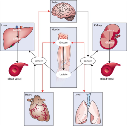
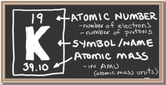
CRIT CLOUD

  • Summaries & Reviews
    • Airway
    • Cardiovascular
    • Controversies
    • Endocrinology
    • Fluids
    • Guidelines
    • Infections
    • Neurology
    • Nutrition
    • Pharmacology
    • Procedures
    • Radiology
    • Renal
    • Resuscitiation
    • Respiratory
    • Sedation
    • Sepsis
    • Transfusion
  • World of Foam
  • Education
    • Download Presentations
    • Download CME Galway Clinic 🔒
    • Download CME Lindenhof Hospital 🔒
    • Echo 🔒
    • Presentations🔒
    • Multimedia
  • About Us
    • Contact

Reviews and Summaries

Out-of-Hospital Cardiac Arrest: The Power of Adrenaline and Amiodarone

29/7/2016

 
Picture
For the resuscitation out-of-hospital one of the mainstays besides compression and defibrillation ist the application of adrenalin and amiodarone. According to the new ACLS guidelines 2015 these are the only drugs remaining in the treatment for shockable rhythms.

​While adrenaline is given for maximum vasoconstriction in order to promote coronary perfusion pressure CPP, amiodarone and sometimes lidocaine are used to promote successful defibrillation of shock-refractory ventricular fibrillation VF or pulseless ventricular tachycardia VT. While the usage of these drugs is undoubtedly very effective in patients with existing circulation the effectiveness during resuscitation remains a matter of debate.

The Effect of Adrenaline

As a matter of fact it has never been proven that adrenalin actually improves long-term outcome. In 2014 Steve Lin and colleagues published a systemativ review on the efficacy of adrenaline in adult out-of-hospital cardiac arrest (OHCA). They were able to show that according to current evidence standard dose adrenaline (1mg) improved rates of survival to hospital admission and return of spontaneous circulation (ROSC) but had no benefit in means of survival to discharge or neurologic outcomes.

What about Amiodarone and Lidocaine?


Kudenchuck et al. now made the effort to look into the efficacy of amiodarone and lidocaine in the setting of OHCA. Used according to the ACLS guidelines 2016 amidarone is given after the third shock applied when treating a shockable rhythm. Two rather small controlled trials have shown so far that using amidarone actually does increase the likelihood of ROSC and the chance to arrive at a hospital alive. It's impact on survival to hospital discharge and neurologic outcome though remains uncertain.

In this randomized, double-blind trial, the investigators compared parenteral amiodarone, lidocaine and saline placebo in adult, non-traumatic, OHCA. They ended up with 3026 patients meeting inclusion criteria and which were randomly assigned to receive amiodarone, lidocaine or saline placebo for treatment. They finally found that neither amiodarone nor lidocaine improved rate of survival to discharge or neurologic outcome significantly. There were also no differences in these outcomes between amiodarone and lidocaine. Across these trial groups also in-hospital care like frequency of coronary catheterisation, therapeutic hypothermia and withdrawal of life-sustaining  treatments did not really differ, making a bias due to treatments after admission unlikely.

Take Home

- This study was not able to show any benefit of amiodarone or lidocaine in the the setting of OHCA  in terms of survival to hospital discharge and neurologic outcome

- Amiodarone seems to improve the likelihood of ROSC and survival to hospital admission (similar to adrenaline)

- As there are no other options, I believe amiodarone should remain part of the standard treatment for shockable rhythms in OHCA

- Lidocaine can be safely removed from CPR sets as there is no benefit of over amiodarone

​
Read here:

N Engl J Med 2016;374:1711-22

Resuscitation, June 2014, Vol 85, Issue 6, p 732-740


New ACLS Guidelines 2015, The Changes

From Review to Practical Guidance on How to Use Ketamine in the ICU

24/6/2016

 
Picture
As posted on BIJC before, Asad et al. had performed a systematic review on the usage of ketamine as a continuous infusion (>24h) in intensive care patients. The same authors have now published a narrative review providing a more depth discussion about the pharmacological and pharmacokinetic properties of ketamine. Also they present recommendations for dosing and monitoring in an ICU setting.

The Goodies of Ket

Current evidence shows that Ketamine... 

- Has no adverse effects on the gastrointestinal tract (bleeding) and does not cause acute kidney injury (compared to nonsteroidal anti-inflammatory drungs, NSAID's) 

- Does not negatively influence bowel motility (in contrast to opioids)

- Preserves laryngeal protective reflexes

- Lowers airway resistance

- Increases lung compliance

- Is less likely to cause respiratory depression

- Is sympathomimetic, facilitates adrenergic transmission and inhibits synaptic catecholamine reuptake, therefore increasing heart rate and blood pressure

The Concerns of Ket

Ketamine...

- Might increase pulmonary airway pressure and therefore aggravate pulmonary hypertension

​- Might cause well known psychotomimetic effects which are of concern in the critically ill patient as this might predispose to delirium

- Interacts with benzodiazepines via the P450 pathway which could result in drug accumulation and prolonged recovery
​

Concerns Proven Wrong

- Ketamine need not to be avoided in patients at risk for seizures, particularly when used for analgosedation for short periods in the ICU setting

- Current evidence shows no increased intracranial pressure or associated adverse neurologic outcomes associated with ketamine administration in critically ill patients
​

Take Home 

The use of ketamine for analgosedation in the ICU continues to lack high-level evidence.However, it is effectively used around the globe and remains an attractive alternative agent for appropriately selected patients. Taking current knowledge and evidence into account this is especially true for patients with severe pain unresponsive to conventional therapies.

Taking precautions and contraindications into account ketamine is considerably safe and even avoids potentially adverse side effects of other agents used.


Erstad BL, J Crit Care, Oct 2016, Vol 35, p 145-149
​

Continuous Etomidate Suppresses the Adrenal Gland in a Dose-Dependent Manner - A Potentially Life-Saving Intervention

4/6/2016


The Problem

Picture
An endogenous Cushing's syndrome, mostly caused by an adenoma of the pituitary gland, is associated with significant morbidity and mortality when left untreated. The condition is closely associated to life-threatening infections, diabetes mellitus, hypertension and increased risk associated with surgery.

For Cushing's disease the first line therapy is surgical removal of the pituitary tumor. Sometimes though urgent medical therapy is needed first. It has been shown, that surgical risk may be significantly reduced if cortisol concentrations are normalised preoperatively. Conditions requiring urgent cortisol-lowering measures are severe biochemical disturbances (e.g. hypokalaemia), immunosuppression or mental instability.

Medical Treatment Options

​Ketokonazole (yes, the antifungal agent) and metyrapone are used to suppress adrenal steroidogenesis at enzymatic sites. Both agents carry the risk of postential side effects. Mifepristone, a glucocorticoid receptor antagonist, and pasireotide, a new targeted pituitary therapy, are alternative agents. However, they also have their limits and side effects.

Etomidate

​Now that's where etomidate joins the game. Interestingly, etomidate and ketokonazole are chemically closely related... they are both members of the imidazole family. Etomidate is used as an anaesthetic agent since 1972 and became popular for hemodynamic stability and the lack if histamine release. In 1983 a Lancet article noted an increased mortality when etomidate was used in critically unwell patients. In 1984 an article in Anaesthesia first showed a link to low serum cortisol levels caused by etomidate. Until now the discussion continues, whether a single induction dose actually negatively influences patient outcome. A meta-analysis in 2010 was unable confirm this apprehension and the debate continues.

Fact is

Etomidate suppresses the production of cortisol by inhibiting the mitochondrial cytochrome p450-dependent adrenal enzyme 11-beta-hydroxylase and therefore lower serum cortisol levels within 12 hours. In higher doses it also blocks side chain cleavage enzymes and also aldosterone synthase. It might even have anti-proliferative effects on adrenal cortical cells.
​On this basis the idea arose, that etomidate might be a useful therapy for severe hypercortisolaemia.

Continuous Etomidate - What's the Evidence

A review article by Preda et al. in 2012 identified 18 publications about the primary therapeutic usage of etomidate in Cushing's syndrome, most of which were case reports. Review of current literature reveals that etomidate indeed suppresses hypercortisolaemia safely and efficiently in patients requiring parenteral therapy. Moreover, etomidate shows a dose-dependent suppression and allows adjustment of the medication to target cortisol levels. At recommended dosages etomidate is considered safe with almost no serious side effects.

The authors conclude, that etomidate is a useful therapeutic option in a hospital setting when oral therapy is not tolerated or inappropriate.
​

Take home

- Continuous etomidate (in non-hypnotic doses) reduces cortisol concentrations in a dose-dependent manner in both hyper- and eucortisolaemic subjects

- The application of continuous etomidate in Cushing's disease is safe and efficient

- After termination of infusion adrenocortical suppression persists for about 3 hours
​
- The suspicion, that a single dose of etomidate for rapid sequence inductions might negatively influence patient outcome in the critically ill remains a matter of debate

​

J Clin Endocrinol Metab. 
1990 May;70(5):1426-30.

Preda et al. European Journal of Endocrinology (2012) 167 137-143       OPEN ACCESS

Soh et al. Letter to the Editor, European Journal of Endocrinology (2012) 167 727–728

Ge et al. Critical Care201317:R20     
OPEN ACCESS

Picture

Lactate - From Bad to Good? An Explanation Trial

14/2/2016

 
Picture
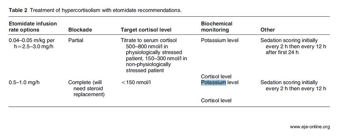
The discussion on the so-called lactic acidosis and its causes have become increasingly attractive over the last couple of years as several biochemical explanations are challenged. A significant confusion persists on the various relationships between lactate, lactic acid and metabolic acidosis. 

Most clinicians continue to refer to the traditional understanding of impaired tissue oxygenation causing increased lactate production, impaired lactate clearance and therefore resultant metabolic acidosis. Just recently we had a discussion on our ward round on this topic when a team member presented the most recent article of UpToDate online on the causes of lactic acidosis. The authors state that 'Lactic acidosis is the most common cause of metabolic acidosis in hospitalised patients' and that 'Lactic acidosis occurs when lactate production exceeds lactate clearance. The increase in lactate production is usually caused by impaired tissue oxygenation...'... finally suggesting that lactate is no good!

These statements support the classical understanding that:
- Hyperlactatemia is caused by tissue hypoxemia, and
- This in turn then leads to a metabolic acidosis called lactic acidosis


This biochemical understanding has persisted for decades, but there are some good reasons to vigorously challenge this traditional aspect on the 'bad' lactate. Lactate turns out to be by far more complex in its characteristics and functions, so I decided to try and make a short but comprehensive overview of this molecule.

What is lactate?

Picture
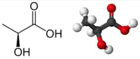
Lactate is a small organic molecule with the chemical formula CH3CH(OH)CO2H and structurally looks like on the image to the left. It is produced in the cytoplasm of human cells mainly by anaerobic glycolysis by the conversion of pyruvate to lactate by LDH. This chemical reaction results typically in a blood lactate to pyruvate ratio of about 10:1. And while lactate is produced, NAD+ also is incurred, and this actually can accept protons itself, so does not result in acidosis itself.

Lactate arises from the production of energy by consuming glycogen and glucose.

Picture


​Where does it come from?

Picture
Typically most people think of muscles first as an origin of lactate. As a matter of fact lactate originates from many other organs, including our red blood cells. Red blood cells always produce lactate as they lack the mitochondria required to regenerate NAD+ needed for glycolysis.  In general, you can say that tissues with lots of LDH are the primary producers of lactate. Around 20mmol/kg/day of lactate are produced under normal circumstances.

Lactate is not only produced in skeletal muscle.

Muscle: 25%
Skin: 25%
Brain: 20%
RBC: 20%
Intestine: 10%

What happens with it?

Lactate is not just for nothing. After its production by anaerobic glycolysis lactate is reutilised, for instance in the liver and the cortex of the kidneys. As an example: under the influence of cortisol it is used for gluconeogenesis in hepatocytes and restores glucose and glycogen. Also, it is a part of oxidative phosphorylation in the liver, kidney, muscles, the heart and the brain. Like this lactate helps conserve glucose levels in our blood.
​
​Lactate actually serves as a fuel for oxidation and glucose regeneration and therefore is a source for energy itself.
Picture
From The Lancet Endocrinology 2013


​
​How does hyperlactatemia develop?

In general, you can assume that there is a balance between lactate production and its consumption or usage. The classical understanding that tissue hypoxia leads to overproduction and underutilisation by impaired mitochondrial oxidation is correct.

The critical point though is that lactate is also produced via aerobic glycolysis as a response to stress. This happens in septic patients, asthmatic exacerbations, trauma and other critical conditions. In these situations, the trigger for lactate production is adrenergic stimulation and NOT tissue hypoxia. There are also several other reasons for hyperlactatemia other than tissue hypoxia:


Sepsis:      Adrenergic drive
Asthma:    Adrenergic drive
Trauma:    Adrenergic drive
Cardiogenic and haemorrhagic shock: Adrenergic drive
Pheochromocytoma: Adrenergic drive
Inflammation: Cytokine drive
Alkalosis, antiretroviral medication and others


Also, there is good evidence showing that organs like the lungs are an important producer of lactate during stress. And of course in all these conditions hypoxic and non-hypoxic hyperlactatemia might also co-exist.

In critically ill patients often other reasons than tissue hypoxia are responsible for hyperlactatemia (e.g. adrenergic drive).
​

Is lactate harmful?

In contrast to the classical understanding of lactate and lactic acidosis more and more evidence comes up indicating that lactate during stress actually serves as a fuel for energy production. Various tissues, e.g. the myocardium increase their lactate uptake during stress significantly. Also, our brain consumes more lactate during stress which is used for oxidation. Research has shown that lactate infusions improve cardiac output in pigs and even in patients with heart failure. 

Experimental work on isolated muscles suggests that circulating catecholamines and development of acidic conditions during exhaustive exercise may improve muscles' tolerance to elevated K+ levels. This implies that during high-intensity activity with high extracellular K+
 and adrenaline, lactate serves as a performance-enhancing chemical, rather than being the cause of muscle fatigue.

Lactate is not harmful to our organism. On the contrary, recent compelling evidence suggests that lactate might be beneficial, rather than detrimental, during high-intensity activity and to force development in working heart and skeletal muscle.
​

Why do critically ill patients with hyperlactatemia die more often then?

In critical care hyperlactatemia indeed is a marker of illness severity and a strong indicator of mortality. This is especially true for patients with sepsis. However, as described above, hyperlactatemia often doesn't indicate hypoperfusion or tissue hypoxia. Hyperlactatemia rather reflects the severity of illness by representing the degree of our body's activation to stress. A fall in lactate concentration following treatment of critically ill patients is due to an attenuation of the stress response rather than to correction of oxygen debt.

​Hyperlactatemia reflects a severe disease and the patients' response to stress. Patients die due to their illness, not because of high lactate.
​

What about Ringer's lactate?

Picture

Ringer's lactate (RL) is not harmful in patients with hyperlactatemia.

As a matter of fact RL turns out to be superior compared to normal saline in hyperlactatemia, acidotic patients and patients with hyperkalemia.
​

The bottom line

- Lactate is an indicator of stress, a marker of illness severity and a strong predictor of mortality, but not harmful as a molecule itself.

- Lactate is helpful
 as an essential source of energy and an important fuel for oxidation and glucose generation.

- During conditions like septic shock, there is no proof that lactate is produced only due to tissue hypoxia. In fact, well-ventilated lungs provide a large amount of lactate during sepsis. Lactate in sepsis and other critical conditions is mostly not due to hypoxemia or hypoperfusion.

- Ringer's lactate contains sodium lactate, but not lactic acid. Lactate itself, as mentioned above, is beneficial in severe disease. Therefore RL remains the fluid of choice during severe disease like for instance septic shock.

- Ringer's lactate is superior to normal saline in patients with metabolic acidosis, hyperlactatemia and also hyperkalemia.
​

Got interested in some better understanding? START READING HERE:

Emmettt et al. UpToDate online, August 2015, Causes of lactic acidosis

Garcia-Alvarez et al. Critical Care 2014, 18:503


Marik PE, Bellomo R. OA Critical Care 2013 Mar 01;1(1):3

Garcia-Alvarez et al. Lancet Diabetes Endocrinol. 2014 Apr;2(4):339-47.

Andersen JB et al. Journal of Experimental Biology  
2007  210: vii doi: 10.1242/jeb.001107​

Bakker J et al. Intensive Care Med (2016) 42:472–474


Also, have a listen to Bellomo's review on lactate:
Picture
Click in image to listen to podcast

Dexmedetomidine vs Midazolam for the Intubated

12/2/2016

 
Picture
Dexmedetomidine has shaken up the usual sedatives in ICU but remains a matter of debate among intensivists. One question is whether the higher costs compared to midazolam are justified by clinical advantages. There is research available suggesting that dexmedetomidine might be an attractive alternative to standard sedatives especially in regards of time to extubation and costs (Turinen et al., Jacob et al.). This seems to hold true for moderate to light sedation of intubated patients.

I've stepped over this prospective, double-blind, randomised trial by Riker et al. in which 68 centres in 5 countries recruited intubated 366 patients to received moderate to light sedation with either dexmedetomidine or midazolam. All patients received daily arousal assessment. 

Their primary end point was the percentage of time within the target sedation range (RASS score −2 to +1) and this did not differ between the two groups.

Looking at the secondary endpoints though make things a lot more interesting. Just before the beginning of the 
sedation period both groups had a similar prevalence of delirium. During study drug administration though, the effect of dexmedetomidine treatment on delirium was significant. A reduction of 24.9% with dexmedetomidine is rather impressive (see figure below). This effect was even greater in patients who were CAM-ICU-positive at baseline.

Finally patients on dexmedetomidine had shorter time to extubation (1.9 days in average) while their length of stay on ICU did not differ.

From a safety point of view the most common adverse effect of dexmedetomidine was bradycardia. It's noteworthy that patients on midazolam had more episodes of hypotension and tachycardia.

THE BOTTOM LINE

- This is another study indicating that dexmedetomidine seems to be beneficial in regards of delirium in mechanically ventilated patients and might speed up time to extubation

- Dexmedetomidine is safe in patients where moderate to light sedation is the aim


Riker et al. JAMA. 2009;301(5):489-499. doi:10.1001/jama.2009.56     OPEN ACCESS


Read more HERE on BIJC

Picture

What is Better in ARDS: Pressure Controlled or Volume Controlled Ventilation?

28/12/2015

 
Picture
A good question, but do you actually know. Most ICU's have their standard modes of ventilation and we are busy enough concentrating on the wright PEEP, the perfect tidal volume or prone positioning the patient. But does the mode of ventilation actually have an impact on the outcome? Chacko et al. had a look at exactly this question and performed a systematic review on this topic:

- Early mortality: There is only some moderate-quality evidence suggesting that pressure controlled ventilation might be of benefit, although this was not observed in the long term follow-up!

- Duration of mechanical ventilation: no apparent difference between pressure- and volume-controlled ventilation

- ICU length of stay: 
no apparent difference between pressure- and volume-controlled ventilation

- incidence of barotrauma: 
no apparent difference
 between pressure- and volume-controlled ventilation

- Extrapulmonary organ failure: One underpowered study in favour of pressure controlled ventilation

- Infective complications, Quality of life: To this date no studies available

Conclusion: Current evidence shows no difference between pressure controlled and volume controlled ventilation in ARDS.


​

Cochrane, Clinical Answers      OPEN ACCESS

Chacko B, Peter JV, Tharyan P, John G, Jeyaseelan L. 
Cochrane Database of Systematic Reviews 2015, Issue 1. Art. No.: CD008807.     OPEN ACCESS

Difficult Airway Society DAS: New Guidelines OUT! Cricoid Pressure still IN?

7/12/2015

 
Picture
November 2015, the Difficult Airway Society have published their updated guidelines for management of difficult intubation in adults. Again DAS provide an excellent overview on unanticipated difficult intubations in adults... worth reading for anyone involved in critical care!

​When reading this article I couldn't help myself putting a special focus on the controversial issue of cricoid pressure (CP) for rapid sequence induction (RSI). This topic has become a major matter of debate as scientific evidence of its effect on preventing aspiration of gastric content is basically lacking. There is quite some evidence available showing that cricoid pressure might actually impair intubation or potentially harm the patient. More background information and links on this topic you can find here
. While some guidelines have actually 'softened' or abandoned the recommendation for the use of CP, most of them have not... and continue to recommend CP. It was therefore of great interest to see what the panel of the DAS would come up with!

For anaesthetists working in Britain and Ireland the DAS guidelines are of special interest as they represent some sort of legal binding on how to proceed at their daily work. We took a closer look at the new guidelines... and got surprised:


"This (CP) is a standard component of rapid sequence induction in the UK". Ok... so no change there! This statement is pretty clear and leaves no space for interpretation - sounds imperative. A little less clear are the following text passages on why CP remains a standard component.

"It is often overlooked that cricoid pressure has been shown to prevent gastric distension during mask ventilation and was originally described for this purpose"... Well, actually cricoid pressure was originally described by Brian Arthur Sellick in the Lancet in 1961 as a preliminary report of an un-controlled case study and the purpose of cricoid pressure was to control regurgitation of gastric content during induction of anaesthesia. 

Picture

​CP and Gastric Insufflation


The reference cited in the context of gastric insufflation and cricoid pressure is an article by Salem and Sellik form Anaesthesia and Analgesia in 1974 (read the original article here). They write: one aim of CP is the prevention of aspiration. The other one is the prevention of gastric insufflation during mask ventilation. The presented evidence in that regard though is not really convincing:
a) A historical letter of Dr. William Cullen... dated back in 1774!
b) The other reference is an article by Salem himself on the 'efficacy of cricoid pressure in preventing gastric inflation during bag-mask ventilation
in paediatric patients, but not adults!

Another article cited by the DAS (
Obstetric Anaesthetists' Association/Difficult Airway Society difficult and failed tracheal intubation guidelines – the way forward for the obstetric airway Br. J. Anaesth. (2015) 115 (6): 815-818) actually recommends gentle ventilation with low insufflation pressure during RSI which should not overcome correctly applied cricoid pressure. This suggests that CP makes gentle bag-mask ventilation safe.
Indeed, 
Lawes et al. already showed in 1987 that when bag-mask ventilating, it was not possible to cause gas to enter the stomach in any patient with a patent airway when cricoid pressure was applied. BUT he also stated that:  In the absence of cricoid pressure the lungs of all the patients could be ventilated “gently” satisfactorily by hand without gas entering the stomach.

​
The Bottom Line

Going through these overall brilliant guidelines by the DAS I still haven't been convinced about the usefulness of cricoid pressure and resume (once again):

- Cricoid pressure for rapid sequence induction remains a non-evidence-based manoeuvre and should be seriously questioned!

​
And by the way, I feel the DAS actually knows that. You have to acknowledge what Hagberg writes in the BJA editorial:
..."the application of CP during rapid sequence induction remains a matter of debate; some believe in its effectiveness in preventing pulmonary aspiration, whereas others believe it should be abandoned because of the paucity of scientific evidence of benefit and possible complications." 
..."The literature does demonstrate that the use of CP is likely to make airway interventions, such as mask ventilation, SGA insertion, direct laryngoscopy, and intubation more difficult."

..."As a result of the lack of sufficient scientific evidence that CP reduces regurgitation, in addition to evidence that it may interfere with airway management..."

​
Any comments?


​
Difficult Airway Society DAS 2015 guidelines for management of unanticipated difficult intubation in adults, Br. J. Anaesth. 2015    OPEN ACCESS

BIJC post on Cricoid Pressure 04/2014

Hagberg et al. DAS 2015 Guidelines - Editorial, Br. J. Anaesth. 2015, 1-3   OPEN ACCESS

Lawes et al. Inflation Pressure, Gastric Insufflation and Rapid Sequence Induction, Br. J. Anaesth. 1987


Spreading the Word: In Hyperkalemia Ringer's Lacate is Superior to Normal Saline

17/10/2015

 
Picture
Just recently the discussion came up once again on what sort of infusion should be used in patients with hyperkalemia. To my surprise the idea seems to persist that normal saline (NS) should be used, as this solute does not contain any further potassium. This is a thought in the wrong direction and Pulmcrit made a great statement in 2014 to clarify this myth. The key points are as follows:

  • Infusing Ringer's lactate (RL) in a patient with hyperkalemia will actually lower his serum potassium level
  • Even a solute with twice the potassium concentration of RL (this would be 8mmol/L) would require a vast amount of fluid to create any effect in serum potassium levels
  • NS has been shown to produce non-anion gap metabolic acidosis, which causes potassium to shift out of cells, thereby increasing potassium levels
  • RL does not cause any acidosis

Here's all the background reading including references:

Pulmcrit Myth-busting: RL is safe in hyperkalemia, and is superior to NS

​
This might also be of interest. Have a very close look on normal saline infusions:

Normal Saline and Acidosis: Is it Really the Salt that Matters?

​

Antiemetics in the Emergency Department: Sure Look it!

18/9/2015

 
Picture
Many patients admitted to the emergency department (ED) suffer of nausea and vomiting - and many doctors treat this with antiemetics like metoclopramide or ondansetron. Treating nausea is tricky and of course we all try to do our best to comfort patients as good as possible. But are you really sure giving an antiemetic in the ED actually improves symptoms?

Unfortunately results coming in on this topic do not look very promising. 3 publications looked at exactly this setting and although their number of patients isn't overwhelming the results are rather discouraging.

Egerton-Warburton and colleagues performed a RCDT and looked a total 258 patients who got either metoclopramide, ondansetron or normal saline as a treatment for nausea in the ED. They basically found no differences in reduction of nausea severity.

Back in 2006 Braude et al. already stated in a RCDT including 97 patients that metoclopramide and prochlorperazine were not more effective than saline placebo as an antiemetics in the ED. Only droperidol was found to be more effective than metoclopramide or prochlorperazine but caused more extrapyramidal symptoms.


And in 2011 Barrett and colleagues published a study with 163 patients where they compared metoclopramide, ondansetron, promethazine and saline placebo in the ED. Same again: no evidence was found that ondansetron is superior to metoclopramide and promethazine in reducing nausea in ED adults.


Even if the number of patients is not that big... it's three trials so far and they all don't really support the use of antiemetics in the emergency department.


It is interesting to note that these drugs have been proven to be effective in the setting of chemotherapy and in anaesthetics, but the setting in the ED seems to differ. At least most patients experienced some relief over time... most probably to treatment of the cause itself!


Egerton-Warburton et al. Ann Emerg Med. 2014;64:526-532      OPEN ACCESS

Braude, D et al. Am J Emerg Med. 2006; 24: 177–182

Barrett et al. Am J Emerg Med. 2011; 29: 247–255


Hemoglobin is NOT Different from Hematocrit... Once and for All!

26/8/2015

 
Picture
It starts in medical school, regularly appears in your medical training, sneaks around nursing schools and is an impetus for discussions in the ICU: The great myths about Hemoglobin (Hb) and Hematocrit (Hct). 

These two haematological lab-parameters are part of our daily life at work and are mostly measured together... as a package. Some clinicians look at haemoglobin levels, others prefer hematocrit levels... but then there is always someone making a great deal of differentiating between the two parameters and making all sort of diagnostic conclusions. 'Hct is better to determine dilution of the patient' or 'Acute blood loss is better determined by Hb than Hct'... and so on.

So here's the question: What actually is the difference between Hb and Hct? Do we need to measure both in clinical practice?


What's the difference?

Hemoglobin levels are mostly measured by automated machines designed to perform different tests in blood. Within the machine, the red blood cells are broken down to get the haemoglobin into a solution. The concentration of haemoglobin is then measured by spectrophotometry using the methemoglobin cyanide method.

Hematocrit levels in contrast are actually calculated by an automated analyzer... It is actually not measured directly! The analyser multiplies the red blood cell count by their mean corpuscular volume.


What is Fact?

There simply is NO difference between Hemoglobin and Hematocrit by means of clinical information!
  • In fact, virtually all haemoglobin in our blood is contained within erythrocytes
  • Therefore, whether the amount of Hb per litre of blood is determined or the blood’s volume occupied by the Hb filled erythrocytes is determined, similar information is gained.
  • Nijboer at al. have brilliantly proven that Hb and Hct correlate in all ranges and all patients and also nicely show this in their figure 1 (see below)
  • The only rare exceptions are macrocytic and polycytemic anaemia in which the Hct is defined by erythrocytes containing a normal mean corpuscular Hb concentration

Conclusion
  • Hemoglobin is NOT different from Hematocrit
  • Both parameters provided identical clinical information


                                                 Once and for all!


Nijboer J et al. J Trauma. 2007;62(5):1310-2.

Click here to read
File Size: 164 kb
File Type: pdf
Download File

Picture

Don't Throw Away your Cooling Devices!

20/8/2015

 
Picture
When performing a kidney transplantation nowadays up to 50% of recipients developed a delayed graft function which is defined as the need of dialysis within seven days. The authors of this recently published NEJM-article asked themselves whether mild hypothermia might influence outcome in this regard. 

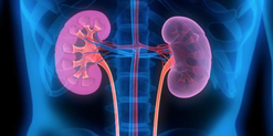
In order to answer this question the investigators assigned organ donors after declaration of death according to neurologic criteria into two groups. They were either treated with mild hypothermia (34 to 35°C) or with normothermia (36.5 to 37.5°C). The target temperature was maintained until the patients were transferred to theatre for transplantation. 

Primary outcome of this trial was delayed graft function among recipients. Secondary outcomes  included the rates of individual organs transplanted into each treatment group at the total number of organs transplanted from each donor. 

This trial had to be stopped early as an interim analysis showed significant efficacy of mild hypothermia. Up to this point a total of 572 patients received a kidney transplant (285 in the hypothermia group and 287 in the normothermia group). 28% of recipients in the hypothemia group developed delayed graft function compared to 39% in the normothermia group.

The authors therefore conclude that mild hypothermia significantly reduces the rate of delayed graft functions among recipients. 

  • This study suggests that potential organ donors after declaration of death according to neurologic criteria should be treated with mild hypothermia. 

  • Intensive care units that continue to treat patients with mild hypothermia after cardiac arrest might have two rewarm their patients for the diagnosis of neurological death before re-cooling them for organ transplantation

Anyhow, it seems reasonable not to get rid of your cooling devices!


Picture
Read more about the controversies of hypothermia in the ICU:


The Targeted Temperature Management Trial: Nielsen N, et al. New Engl J Med. 2013 Dec;369(23):2197-206

The 2 trials that introduced therapeutic hypothermia into ICU practice:
The Hypothermia After Cardiac Arrest Study Group, Holzer at al. New Engl J Med. 2002 Feb;346(8):549-556

Bernard S.A. et al. New Engl J Med. 2002 Feb;346(8):557-563

Review article on therapeutic hypothermia for non-VF/VT cardiac arrest:
Sandroni S. et al. Crit Care Med; 2013;17:215

Pyrexia and neurological outcome:
Leary M. et al. Resuscitation. 2013 Aug;84(8):1056-61


BIJC post on: The Effect of Pre-Hospital Cooling: Rather Worrying Results


Another Hole in the Ballon (-Pump)!

7/5/2015

 
Picture
In the late 1960's the technology of counter-pulsation by using an intra-aortic balloon pump (IABP) was introduced into clinical work. Based on the principle of diastolic inflation and systolic deflation, IABP counter-pulsation improves diastolic coronary artery blood flow and decreases left ventricular afterload. Up to the year 2009, 2012 respectively, the usage of an IABP in patients with ST-segment elevation myocardial infarction and cardiogenic shock was considered a class IC recommendation (reminder: levels of evidence).

Since then a couple of well conducted, larger trials have failed to show a positive impact of IABP especially on mortality. In regards of the most recent meta-analysis in JAMA we provide a short overview of the most important publications. It's interesting to see that the balloons undermining started with a meta-analysis and for the the time being ends with one.


Stitch no.1

The first notable hole in the ballon was caused by Sjauw et al.'s systematic review and meta-analysis in the European Heart Journal in 2009. Their pooled randomized data consisting of two separate meta-analyses did not support the use of an IABP in patients with high risk STEMI. They concluded that
there is insufficient evidence endorsing the current guideline recommendation for the use of IABP therapy in the setting of STEMI complicated by cardiogenic shock.

This publication was one of the main reasons for the expert panel of the European Society of Cardiology to change the recommendation (ESC Guidelines 2012) to use an IABP in patients with STEMI from IC to IIB.


Stitch 2 and 3

In the same year 2012 Thiele et al. published their first IABP-SHOCK II results in the NEJM. Their

randomized, prospective, open-label, multicenter trial showed no reduction in the 30-day mortality
compared to the best available medical therapy alone in patients with myocardial infarction-induced cardiogenic shock and planned early revascularization (PCI or CABG).

One year later the IABP-SHOCK II investigators published their final 12-months results in The Lancet. They came to the final conclusion that in patients undergoing early revascularization for myocardial infarction with cardiogenic shock, IABP did not reduce 12-month all-cause mortality.


Stitch no. 4

In 2013 Ranucci at al. presented the results of their
single-center prospective randomized controlled trial looking at the usage of a preoperative IABP in high-risk patients undergoing surgical coronary revascularization. By looking at a total of 110 patients with an ejection fraction below 35% and no hemodynamic instability there was no improvement in outcome when inserting an IABP preoperatively.

Preliminary Final Stitch

So finally Ahmad and his team decided to assess IABP efficacy in acute myocardial infarction by performing an updated meta-analysis. Main outcome was 30-day mortality. They included
12 eligible RCTs randomizing 2123 patients and found no improvement in mortality among patients with acute myocardial infarction... regardless of whether patients had cardiogenic shock or not!
A look at another 15 eligible observational studies with a total of 15 530 patients showed basically conflicting results which was explained by the differences between studies in the balance of risk factors between IABP and non-IABP groups.


It seems that the IABP fails to show its assumed efficacy in patients with myocardial infarction and cardiogenic shock, especially when early revascularization (PCI or CABG) is available.

As a general consideration and also when no early revascularisation is available the use of another left-ventricular assist device like the Impella pump might prove to be a good and easy to use alternative (see blow).


Sjauw KD et al. Eur Heart J 30: 459-468

ESC Guidelines for the management of acute myocardial infarction in patients presenting with ST-segment elevation. Eur Heart J 33: 2569-2619        
OPEN ACCESS

Thiele et al. N Engl J Med 2012; 367:1287-1296            OPEN ACCESS

Thiele et al. The Lancet, Volume 382, Issue 9905, Pages 1638 – 1645

Crit Care Med. 2013 Nov;41(11):2476-83

JAMA Intern Med. Published online March 30, 2015


Short film on the principle of the Impella pump 2.5. Bare in mind that this device can actually be easily inserted in the environment of ICU and positioned by using transthoracic echo TTE.


Lancet vs Cochrane: ICU's Should Keep Neuraminidase Inhibitors in Stock

6/5/2015

 
Picture
Almost exactly one year ago the Cochrane Library published an intervention review on the prevention and treatment of influenza with neuraminidase inhibitors in adults and children. The reason for this review was the fact that many countries stockpile these drugs and the WHO classified them as an essential medicine.
Jefferson et al. used the data of 46 trials with oseltamivir or zanamivir for this review. They basically conclude that:

- Both drugs shorten the duration of symptoms of influenza-like symptoms by less than a day
- Oseltamivir did not affect the number of hospitalizations
- Prophylaxis trials showed a reduced risk of symptomatic influenza in individuals and households, but no definite conclusion can be made
- Oseltamivir use was associated though with nausea, vomiting, headaches, renal and psychiatric events

...and finally write: 'The influenza virus-specific mechanism of action proposed by the producers does not fit the clinical evidence'. This review certainly undermined the importance of oseltamivir for many of us.


The Cochrane review though did not look at outcomes like mortality, but the Lancet Respiratory Medicine did! Stella G at al. have now published a large systematic review which included 29'234 patients from 78 studies during the period from 2009 to 2014. Their findings come rather surprisingly:

-
Compared with no treatment, neuraminidase inhibitor treatment (irrespective of timing) was associated with a reduction in mortality risk
- Compared with later treatment, early treatment (within 2 days of symptom onset) was associated with a reduction in mortality risk
- The reduction in mortality risk was observed when treatment was started up to 5 days of symptoms onset


There still seem to be some good reasons to use oseltamivir in critically ill patients with suspected or proven influenza... up to 5 days of symptoms onset!


Jefferson T et al. The Cochrane Collaboration,
Published Online: 10 APR 2014

The Cochrane Collaboration News Release 10 April 2014

Muthuri, Stella G et al. The Lancet Respiratory Medicine , Volume 2 , Issue 5 , 395 - 404


As ProMISe'd... the Trial is Complete!

25/3/2015

 
Picture
As every child already knows by now the study by Rivers et al. in 2002 has raised the awareness about sepsis and led to the establishment of the surviving sepsis campaign. As we have posted on BIJC before, many elements of the early goal directed therapy (EGDT) have been discussed controversially since. In order to answer some of the questions of sepsis treatment three big trials have been started, involving different parts of this world. These efforts have led to a unique situation as we now have three high quality trials looking at the classical EGDT versus 'usual care'.

ARISE and ProCESS had been published before (read here) and both of them showed no difference between EGDT and 'usual care'.

ProMISe included 1251 patients with severe sepsis or septic shock that were admitted to a total 56 hospitals in the UK. Again classical EGDT with measurement of continuous central venous oxygenation was compared to so called 'usual treatment'. It's remarkable to notice that in the 'usual treatment' group about half of the patient didn't get a central line and central venous oxygenation wasn't even measured in the ones who got one. And here's the result:
There was no difference in 90-day mortality and no differences in secondary outcomes. In contrast EGDT actually increased costs.

It has become difficult to ignore these three trials!


Our conclusion: The classical EGDT therapy has ended here and now... but EGDT will keep its central role in the treatment of septic patients!

Early:
- Identify septic patient quickly, start screening for patients if indicated
- Administer antibiotics within the first our of recognition of sepsis
- Start IV-fluid therapy immediately
- Take (blood) cultures as quick as possible, but do not delay antibiotic treatment

Goal Directed:
- Aim for a reasonable mean arterial pressure (e.g. 65mmHg)
- Aim for a sufficient urinary output (0.5ml/h)
-
Central venous pressure (CVP) certainly and most probably central venous oxygenation (ScvO2) are not parameters to measure fluid responsiveness
- Lactate remains an issue of debate

Therapy:
- Simple: Whatever the physician feels is best!


ProMISe Trial, Mouncey et al. N Engl J Med. 2015 Mar 17.

BIJC Review on ARISE and ProCESS


Picture displayed taken from the Ice Cream Trilogy by Wright, Pegg and Frost

Doctors Should Position their Name Tag on the Right Side of their Chest... It's Evidence Based!

23/3/2015

 
PictureThey're All WRONG!!!
Here it is finally, the study we all have been waiting for. 'THE question' that has never been answered... until now!

Every day we all do our best to establish the best patient-physician relation possible. The first impression counts and besides introducing ourselves verbally we ware name tags to visually inform about our name and position. But did anyone actually ever ask himself if it makes a difference wether you ware your name tag on the left or right side of your chest?

Well, an orthopaedic surgeon in Switzerland obviously has and conducted a 'blinded' study to answer this specific question. He made one hundred volunteers, blinded to the experimental setup, present for an orthopedic consultation in a standardized manner. The name tag of the physician was randomly positioned on the left chest side and presented to 50 individuals (age 35 years (range 17 to 83)) or the right chest side and then presented to 50 other individuals (35 years (range 16 to 59)). The time of the participant noticing the name tag was documented. Subsequently, the participant was questioned concerning the relevance of a name tag and verbal self-introduction of the physician.


38% of the participants noticed the nametag on the right as opposed to 20% who noticed it if placed on the left upper chest... hey, this turned out to be statistically significant, giving us a p-value of 0.0473!

The author concludes:  Positioning the name tag on the right chest side results in better and faster visibility.

We conclude:
- Also orthopaedic surgeons seem to be interested in a close patient-physician relationship
- Orthopedic surgeons actually do talk to their patients!
- Orthopedic surgeons in Switzerland don't seem to be overstrained with their workload


Schmid SL et al. March 2015, PLOS One, DOI: 10.1371/journal.pone.0119042

<<Previous
Forward>>

    Search


    ​Translate Page

    Select your language above. Beware: Google Translate is often imprecise and might result in incorrect phrases!

    Picture


    ​Categories

    All
    Airway
    Cardiovascular
    Controversies
    Endocrinology
    Fluids
    For A Smile ; )
    Guidelines
    Infections
    Meducation
    Neurology
    Nutrition
    Pharmacology
    Procedures
    Radiology
    Renal
    Respiratory
    Resuscitation
    SARS CoV 2
    SARS-CoV-2
    Sedation
    Sepsis
    Transfusion

    Archives

    December 2025
    June 2025
    January 2021
    September 2020
    March 2020
    February 2020
    January 2020
    December 2019
    November 2019
    July 2019
    May 2019
    March 2019
    February 2019
    January 2019
    December 2018
    January 2018
    October 2017
    August 2017
    June 2017
    March 2017
    February 2017
    January 2017
    October 2016
    July 2016
    June 2016
    April 2016
    February 2016
    December 2015
    October 2015
    September 2015
    August 2015
    July 2015
    June 2015
    May 2015
    April 2015
    March 2015
    January 2015
    December 2014
    November 2014
    October 2014
    September 2014
    August 2014
    July 2014
    June 2014
    May 2014
    April 2014
    March 2014
    February 2014
    January 2014
    December 2013
    November 2013

    Author

    Timothy Aebi

    RSS Feed

ABOUT US

The Crit ☁​ supports free access medical education. The content of this website can be used and reproduced by stating a reference to our site.
Read our statements here

SEARCH US

CONTACT US

If you have any interesting article or news to be posted on this website, please contact us here
​

FOLLOW US

Non-AI-Website

These Blog-Posts are not written by Artificial Intelligence AI. There might be some mistypings or grammatical mistakes - but therefore no AI-writing involved!
Picture