
CRIT CLOUD

  • Summaries & Reviews
    • Airway
    • Cardiovascular
    • Controversies
    • Endocrinology
    • Fluids
    • Guidelines
    • Infections
    • Neurology
    • Nutrition
    • Pharmacology
    • Procedures
    • Radiology
    • Renal
    • Resuscitiation
    • Respiratory
    • Sedation
    • Sepsis
    • Transfusion
  • World of Foam
  • Education
    • Download Presentations
    • Download CME Galway Clinic 🔒
    • Download CME Lindenhof Hospital 🔒
    • Echo 🔒
    • Presentations🔒
    • Multimedia
  • About Us
    • Contact
  • Summaries & Reviews
    • Airway
    • Cardiovascular
    • Controversies
    • Endocrinology
    • Fluids
    • Guidelines
    • Infections
    • Neurology
    • Nutrition
    • Pharmacology
    • Procedures
    • Radiology
    • Renal
    • Resuscitiation
    • Respiratory
    • Sedation
    • Sepsis
    • Transfusion
  • World of Foam
  • Education
    • Download Presentations
    • Download CME Galway Clinic 🔒
    • Download CME Lindenhof Hospital 🔒
    • Echo 🔒
    • Presentations🔒
    • Multimedia
  • About Us
    • Contact

Reviews and Summaries

Lactate - From Bad to Good? An Explanation Trial

14/2/2016

 
Picture
The discussion on the so-called lactic acidosis and its causes have become increasingly attractive over the last couple of years as several biochemical explanations are challenged. A significant confusion persists on the various relationships between lactate, lactic acid and metabolic acidosis. 

Most clinicians continue to refer to the traditional understanding of impaired tissue oxygenation causing increased lactate production, impaired lactate clearance and therefore resultant metabolic acidosis. Just recently we had a discussion on our ward round on this topic when a team member presented the most recent article of UpToDate online on the causes of lactic acidosis. The authors state that 'Lactic acidosis is the most common cause of metabolic acidosis in hospitalised patients' and that 'Lactic acidosis occurs when lactate production exceeds lactate clearance. The increase in lactate production is usually caused by impaired tissue oxygenation...'... finally suggesting that lactate is no good!

These statements support the classical understanding that:
- Hyperlactatemia is caused by tissue hypoxemia, and
- This in turn then leads to a metabolic acidosis called lactic acidosis


This biochemical understanding has persisted for decades, but there are some good reasons to vigorously challenge this traditional aspect on the 'bad' lactate. Lactate turns out to be by far more complex in its characteristics and functions, so I decided to try and make a short but comprehensive overview of this molecule.

What is lactate?
Picture
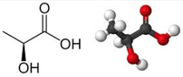
Lactate is a small organic molecule with the chemical formula CH3CH(OH)CO2H and structurally looks like on the image to the left. It is produced in the cytoplasm of human cells mainly by anaerobic glycolysis by the conversion of pyruvate to lactate by LDH. This chemical reaction results typically in a blood lactate to pyruvate ratio of about 10:1. And while lactate is produced, NAD+ also is incurred, and this actually can accept protons itself, so does not result in acidosis itself.

Lactate arises from the production of energy by consuming glycogen and glucose.

Picture


​Where does it come from?

Picture
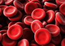
Typically most people think of muscles first as an origin of lactate. As a matter of fact lactate originates from many other organs, including our red blood cells. Red blood cells always produce lactate as they lack the mitochondria required to regenerate NAD+ needed for glycolysis.  In general, you can say that tissues with lots of LDH are the primary producers of lactate. Around 20mmol/kg/day of lactate are produced under normal circumstances.

Lactate is not only produced in skeletal muscle.

Muscle: 25%
Skin: 25%
Brain: 20%
RBC: 20%
Intestine: 10%

What happens with it?

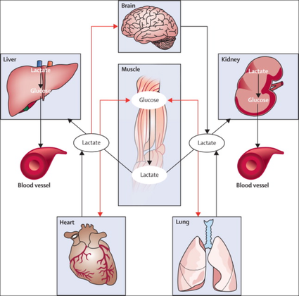
Lactate is not just for nothing. After its production by anaerobic glycolysis lactate is reutilised, for instance in the liver and the cortex of the kidneys. As an example: under the influence of cortisol it is used for gluconeogenesis in hepatocytes and restores glucose and glycogen. Also, it is a part of oxidative phosphorylation in the liver, kidney, muscles, the heart and the brain. Like this lactate helps conserve glucose levels in our blood.
​
​Lactate actually serves as a fuel for oxidation and glucose regeneration and therefore is a source for energy itself.
Picture
From The Lancet Endocrinology 2013


​
​How does hyperlactatemia develop?

In general, you can assume that there is a balance between lactate production and its consumption or usage. The classical understanding that tissue hypoxia leads to overproduction and underutilisation by impaired mitochondrial oxidation is correct.

The critical point though is that lactate is also produced via aerobic glycolysis as a response to stress. This happens in septic patients, asthmatic exacerbations, trauma and other critical conditions. In these situations, the trigger for lactate production is adrenergic stimulation and NOT tissue hypoxia. There are also several other reasons for hyperlactatemia other than tissue hypoxia:


Sepsis:      Adrenergic drive
Asthma:    Adrenergic drive
Trauma:    Adrenergic drive
Cardiogenic and haemorrhagic shock: Adrenergic drive
Pheochromocytoma: Adrenergic drive
Inflammation: Cytokine drive
Alkalosis, antiretroviral medication and others


Also, there is good evidence showing that organs like the lungs are an important producer of lactate during stress. And of course in all these conditions hypoxic and non-hypoxic hyperlactatemia might also co-exist.

In critically ill patients often other reasons than tissue hypoxia are responsible for hyperlactatemia (e.g. adrenergic drive).
​

Is lactate harmful?

In contrast to the classical understanding of lactate and lactic acidosis more and more evidence comes up indicating that lactate during stress actually serves as a fuel for energy production. Various tissues, e.g. the myocardium increase their lactate uptake during stress significantly. Also, our brain consumes more lactate during stress which is used for oxidation. Research has shown that lactate infusions improve cardiac output in pigs and even in patients with heart failure. 

Experimental work on isolated muscles suggests that circulating catecholamines and development of acidic conditions during exhaustive exercise may improve muscles' tolerance to elevated K+ levels. This implies that during high-intensity activity with high extracellular K+
 and adrenaline, lactate serves as a performance-enhancing chemical, rather than being the cause of muscle fatigue.

Lactate is not harmful to our organism. On the contrary, recent compelling evidence suggests that lactate might be beneficial, rather than detrimental, during high-intensity activity and to force development in working heart and skeletal muscle.
​

Why do critically ill patients with hyperlactatemia die more often then?

In critical care hyperlactatemia indeed is a marker of illness severity and a strong indicator of mortality. This is especially true for patients with sepsis. However, as described above, hyperlactatemia often doesn't indicate hypoperfusion or tissue hypoxia. Hyperlactatemia rather reflects the severity of illness by representing the degree of our body's activation to stress. A fall in lactate concentration following treatment of critically ill patients is due to an attenuation of the stress response rather than to correction of oxygen debt.

​Hyperlactatemia reflects a severe disease and the patients' response to stress. Patients die due to their illness, not because of high lactate.
​

What about Ringer's lactate?

Picture

Ringer's lactate (RL) is not harmful in patients with hyperlactatemia.

As a matter of fact RL turns out to be superior compared to normal saline in hyperlactatemia, acidotic patients and patients with hyperkalemia.
​

The bottom line

- Lactate is an indicator of stress, a marker of illness severity and a strong predictor of mortality, but not harmful as a molecule itself.

- Lactate is helpful
 as an essential source of energy and an important fuel for oxidation and glucose generation.

- During conditions like septic shock, there is no proof that lactate is produced only due to tissue hypoxia. In fact, well-ventilated lungs provide a large amount of lactate during sepsis. Lactate in sepsis and other critical conditions is mostly not due to hypoxemia or hypoperfusion.

- Ringer's lactate contains sodium lactate, but not lactic acid. Lactate itself, as mentioned above, is beneficial in severe disease. Therefore RL remains the fluid of choice during severe disease like for instance septic shock.

- Ringer's lactate is superior to normal saline in patients with metabolic acidosis, hyperlactatemia and also hyperkalemia.
​

Got interested in some better understanding? START READING HERE:

Emmettt et al. UpToDate online, August 2015, Causes of lactic acidosis

Garcia-Alvarez et al. Critical Care 2014, 18:503


Marik PE, Bellomo R. OA Critical Care 2013 Mar 01;1(1):3

Garcia-Alvarez et al. Lancet Diabetes Endocrinol. 2014 Apr;2(4):339-47.

Andersen JB et al. Journal of Experimental Biology  
2007  210: vii doi: 10.1242/jeb.001107​

Bakker J et al. Intensive Care Med (2016) 42:472–474


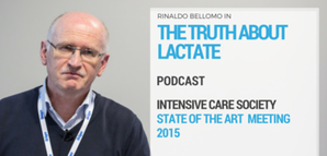
Also, have a listen to Bellomo's review on lactate:
Picture
Click in image to listen to podcast

What is Better in ARDS: Pressure Controlled or Volume Controlled Ventilation?

28/12/2015

 
Picture
A good question, but do you actually know. Most ICU's have their standard modes of ventilation and we are busy enough concentrating on the wright PEEP, the perfect tidal volume or prone positioning the patient. But does the mode of ventilation actually have an impact on the outcome? Chacko et al. had a look at exactly this question and performed a systematic review on this topic:

- Early mortality: There is only some moderate-quality evidence suggesting that pressure controlled ventilation might be of benefit, although this was not observed in the long term follow-up!

- Duration of mechanical ventilation: no apparent difference between pressure- and volume-controlled ventilation

- ICU length of stay: 
no apparent difference between pressure- and volume-controlled ventilation

- incidence of barotrauma: 
no apparent difference
 between pressure- and volume-controlled ventilation

- Extrapulmonary organ failure: One underpowered study in favour of pressure controlled ventilation

- Infective complications, Quality of life: To this date no studies available

Conclusion: Current evidence shows no difference between pressure controlled and volume controlled ventilation in ARDS.


​

Cochrane, Clinical Answers      OPEN ACCESS

Chacko B, Peter JV, Tharyan P, John G, Jeyaseelan L. 
Cochrane Database of Systematic Reviews 2015, Issue 1. Art. No.: CD008807.     OPEN ACCESS

Difficult Airway Society DAS: New Guidelines OUT! Cricoid Pressure still IN?

7/12/2015

 
Picture
November 2015, the Difficult Airway Society have published their updated guidelines for management of difficult intubation in adults. Again DAS provide an excellent overview on unanticipated difficult intubations in adults... worth reading for anyone involved in critical care!

​When reading this article I couldn't help myself putting a special focus on the controversial issue of cricoid pressure (CP) for rapid sequence induction (RSI). This topic has become a major matter of debate as scientific evidence of its effect on preventing aspiration of gastric content is basically lacking. There is quite some evidence available showing that cricoid pressure might actually impair intubation or potentially harm the patient. More background information and links on this topic you can find here
. While some guidelines have actually 'softened' or abandoned the recommendation for the use of CP, most of them have not... and continue to recommend CP. It was therefore of great interest to see what the panel of the DAS would come up with!

For anaesthetists working in Britain and Ireland the DAS guidelines are of special interest as they represent some sort of legal binding on how to proceed at their daily work. We took a closer look at the new guidelines... and got surprised:


"This (CP) is a standard component of rapid sequence induction in the UK". Ok... so no change there! This statement is pretty clear and leaves no space for interpretation - sounds imperative. A little less clear are the following text passages on why CP remains a standard component.

"It is often overlooked that cricoid pressure has been shown to prevent gastric distension during mask ventilation and was originally described for this purpose"... Well, actually cricoid pressure was originally described by Brian Arthur Sellick in the Lancet in 1961 as a preliminary report of an un-controlled case study and the purpose of cricoid pressure was to control regurgitation of gastric content during induction of anaesthesia. 

Picture

​CP and Gastric Insufflation


The reference cited in the context of gastric insufflation and cricoid pressure is an article by Salem and Sellik form Anaesthesia and Analgesia in 1974 (read the original article here). They write: one aim of CP is the prevention of aspiration. The other one is the prevention of gastric insufflation during mask ventilation. The presented evidence in that regard though is not really convincing:
a) A historical letter of Dr. William Cullen... dated back in 1774!
b) The other reference is an article by Salem himself on the 'efficacy of cricoid pressure in preventing gastric inflation during bag-mask ventilation
in paediatric patients, but not adults!

Another article cited by the DAS (
Obstetric Anaesthetists' Association/Difficult Airway Society difficult and failed tracheal intubation guidelines – the way forward for the obstetric airway Br. J. Anaesth. (2015) 115 (6): 815-818) actually recommends gentle ventilation with low insufflation pressure during RSI which should not overcome correctly applied cricoid pressure. This suggests that CP makes gentle bag-mask ventilation safe.
Indeed, 
Lawes et al. already showed in 1987 that when bag-mask ventilating, it was not possible to cause gas to enter the stomach in any patient with a patent airway when cricoid pressure was applied. BUT he also stated that:  In the absence of cricoid pressure the lungs of all the patients could be ventilated “gently” satisfactorily by hand without gas entering the stomach.

​
The Bottom Line

Going through these overall brilliant guidelines by the DAS I still haven't been convinced about the usefulness of cricoid pressure and resume (once again):

- Cricoid pressure for rapid sequence induction remains a non-evidence-based manoeuvre and should be seriously questioned!

​
And by the way, I feel the DAS actually knows that. You have to acknowledge what Hagberg writes in the BJA editorial:
..."the application of CP during rapid sequence induction remains a matter of debate; some believe in its effectiveness in preventing pulmonary aspiration, whereas others believe it should be abandoned because of the paucity of scientific evidence of benefit and possible complications." 
..."The literature does demonstrate that the use of CP is likely to make airway interventions, such as mask ventilation, SGA insertion, direct laryngoscopy, and intubation more difficult."

..."As a result of the lack of sufficient scientific evidence that CP reduces regurgitation, in addition to evidence that it may interfere with airway management..."

​
Any comments?


​
Difficult Airway Society DAS 2015 guidelines for management of unanticipated difficult intubation in adults, Br. J. Anaesth. 2015    OPEN ACCESS

BIJC post on Cricoid Pressure 04/2014

Hagberg et al. DAS 2015 Guidelines - Editorial, Br. J. Anaesth. 2015, 1-3   OPEN ACCESS

Lawes et al. Inflation Pressure, Gastric Insufflation and Rapid Sequence Induction, Br. J. Anaesth. 1987


OUT NOW: New and Updated ILCOR 2015 Treatment Recommendations on Cardiopulmonary Resuscitation

23/10/2015

 
Picture
Since the year 2000 the International Liaison Committee on Resuscitation (ILCOR) continues to evaluate all evidence and updates their recommendations in 5-year cycles. The most recent ILCOR 2015 International Consensus Conference was held in Dallas last February and the new treatment recommendation are out now.

Resuscitation remains one of the most challenging situations in health care. Providing basic and advanced cardiac life support gives you the opportunity to virtually safe a patients life but in a very limited period of time. It is an enormous challenge to consider all emerging evidence and pack this into simple and useful guidelines.

It is imperative to for any health care provider to get familiar with the updated guidelines and major changes. Below you can find all relevant links to get the reading going. 

The team of BoringEM.org in Canada have provided some excellent infographics to visualise all important changes in the new treatment guidelines since 2010. You should also note that the Canadian Heart & Stroke Association and the American Heart Association have just published the 'HIGHLIGHTS of the 2015 American Heart Association Guidelines Update for CPR and ECC', an excellent summary of the new recommendations and changes. So if you can't find the time to read all of the publication in 'Circulation', this will certainly provide all information you need to know.


Summary of the Canadian Heart & Stroke Association and the American Heart Association: HIGHLIGHTS of the 2015 American Heart Association Guidelines Update for CPR and ECC

​
​
The original publication in Circulation, October 20, 2015, Volume 132, Issue 16 suppl 1

OPEN ACCESS


The Most Important Changes (Click to Enlarge)


The Updated Algorithms (Click to Enlarge)
​


​ERC and ESICM 2015 Guidelines for Post-Resuscitation Care
​

​Based on the the 2015 ILCOR treatment recommendations the European Resuscitation Council (ERC) and the European Society of Intensive Care Medicine (ESICM) have produced these post-resuscitation care guidelines on October the 13th. Recent changes here are the greater emphasis for urgent PCI when indicated, target temperature management at 36°C, prognostic evaluation using a multimodal strategy and an increased emphasis on rehabilitation after survival.
​
​
Nolan JP, Resuscitation, October 2015, Pages 202 - 222

​

Spreading the Word: In Hyperkalemia Ringer's Lacate is Superior to Normal Saline

17/10/2015

 
Picture
Just recently the discussion came up once again on what sort of infusion should be used in patients with hyperkalemia. To my surprise the idea seems to persist that normal saline (NS) should be used, as this solute does not contain any further potassium. This is a thought in the wrong direction and Pulmcrit made a great statement in 2014 to clarify this myth. The key points are as follows:

  • Infusing Ringer's lactate (RL) in a patient with hyperkalemia will actually lower his serum potassium level
  • Even a solute with twice the potassium concentration of RL (this would be 8mmol/L) would require a vast amount of fluid to create any effect in serum potassium levels
  • NS has been shown to produce non-anion gap metabolic acidosis, which causes potassium to shift out of cells, thereby increasing potassium levels
  • RL does not cause any acidosis

Here's all the background reading including references:

Pulmcrit Myth-busting: RL is safe in hyperkalemia, and is superior to NS

​
This might also be of interest. Have a very close look on normal saline infusions:

Normal Saline and Acidosis: Is it Really the Salt that Matters?

​

Antiemetics in the Emergency Department: Sure Look it!

18/9/2015

 
Picture
Many patients admitted to the emergency department (ED) suffer of nausea and vomiting - and many doctors treat this with antiemetics like metoclopramide or ondansetron. Treating nausea is tricky and of course we all try to do our best to comfort patients as good as possible. But are you really sure giving an antiemetic in the ED actually improves symptoms?

Unfortunately results coming in on this topic do not look very promising. 3 publications looked at exactly this setting and although their number of patients isn't overwhelming the results are rather discouraging.

Egerton-Warburton and colleagues performed a RCDT and looked a total 258 patients who got either metoclopramide, ondansetron or normal saline as a treatment for nausea in the ED. They basically found no differences in reduction of nausea severity.

Back in 2006 Braude et al. already stated in a RCDT including 97 patients that metoclopramide and prochlorperazine were not more effective than saline placebo as an antiemetics in the ED. Only droperidol was found to be more effective than metoclopramide or prochlorperazine but caused more extrapyramidal symptoms.


And in 2011 Barrett and colleagues published a study with 163 patients where they compared metoclopramide, ondansetron, promethazine and saline placebo in the ED. Same again: no evidence was found that ondansetron is superior to metoclopramide and promethazine in reducing nausea in ED adults.


Even if the number of patients is not that big... it's three trials so far and they all don't really support the use of antiemetics in the emergency department.


It is interesting to note that these drugs have been proven to be effective in the setting of chemotherapy and in anaesthetics, but the setting in the ED seems to differ. At least most patients experienced some relief over time... most probably to treatment of the cause itself!


Egerton-Warburton et al. Ann Emerg Med. 2014;64:526-532      OPEN ACCESS

Braude, D et al. Am J Emerg Med. 2006; 24: 177–182

Barrett et al. Am J Emerg Med. 2011; 29: 247–255


Hemoglobin is NOT Different from Hematocrit... Once and for All!

26/8/2015

 
Picture
It starts in medical school, regularly appears in your medical training, sneaks around nursing schools and is an impetus for discussions in the ICU: The great myths about Hemoglobin (Hb) and Hematocrit (Hct). 

These two haematological lab-parameters are part of our daily life at work and are mostly measured together... as a package. Some clinicians look at haemoglobin levels, others prefer hematocrit levels... but then there is always someone making a great deal of differentiating between the two parameters and making all sort of diagnostic conclusions. 'Hct is better to determine dilution of the patient' or 'Acute blood loss is better determined by Hb than Hct'... and so on.

So here's the question: What actually is the difference between Hb and Hct? Do we need to measure both in clinical practice?


What's the difference?

Hemoglobin levels are mostly measured by automated machines designed to perform different tests in blood. Within the machine, the red blood cells are broken down to get the haemoglobin into a solution. The concentration of haemoglobin is then measured by spectrophotometry using the methemoglobin cyanide method.

Hematocrit levels in contrast are actually calculated by an automated analyzer... It is actually not measured directly! The analyser multiplies the red blood cell count by their mean corpuscular volume.


What is Fact?

There simply is NO difference between Hemoglobin and Hematocrit by means of clinical information!
  • In fact, virtually all haemoglobin in our blood is contained within erythrocytes
  • Therefore, whether the amount of Hb per litre of blood is determined or the blood’s volume occupied by the Hb filled erythrocytes is determined, similar information is gained.
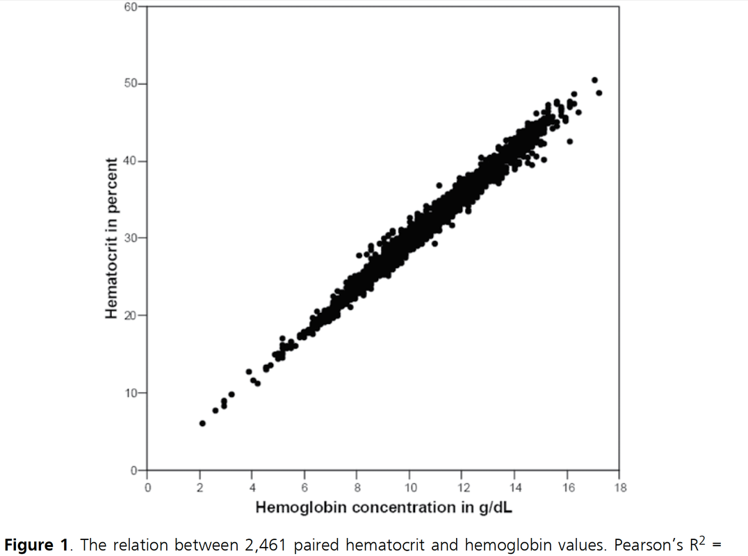
  • Nijboer at al. have brilliantly proven that Hb and Hct correlate in all ranges and all patients and also nicely show this in their figure 1 (see below)
  • The only rare exceptions are macrocytic and polycytemic anaemia in which the Hct is defined by erythrocytes containing a normal mean corpuscular Hb concentration

Conclusion
  • Hemoglobin is NOT different from Hematocrit
  • Both parameters provided identical clinical information


                                                 Once and for all!


Nijboer J et al. J Trauma. 2007;62(5):1310-2.

Click here to read
File Size: 164 kb
File Type: pdf
Download File

Picture

New Guidelines on the Treatment of Idiopathic Pulmonary Fibrosis - Get Updated!

24/8/2015

 
Picture
Idiopathic pulmonary fibrosis is one of these frustrating diseases you repeatedly encounter in the ICU and that mostly leaves you sort of frustrated at the end. Despite all the efforts in research we are still left with very little we can do. This is one reason why also intensivists need to keep themselves updated on this topic. 

As knowledge is growing the ATS, ERS, JRS and ALAT (... thoracic and respiratory societies) made the effort to look into the latest evidence by performing systematic reviews and where appropriate meta-analyses. The aim was to update the guidelines published in 2011. These guidelines are also dedicated to Mr. William Cunningham who actively participated in the development of these guidelines, suffered from idiopathic pulmonary fibrosis for many years and who was directly confronted with the issues related with this condition.


The main conclusions can be briefly summarised as follows:
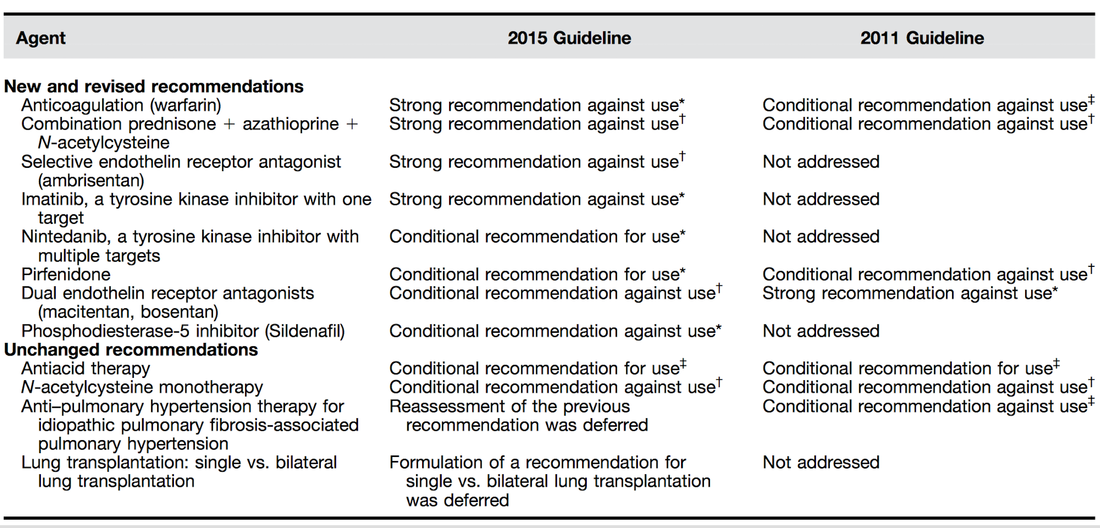
Picture

An Official ATS/ERS/JRS/ALAT Clinical Practice Guideline: Treatment of Idiopathic Pulmonary Fibrosis. An Update of the 2011 Clinical Practice Guideline, American Journal of Respiratory and Critical Care Medicine, Vol. 192, No. 2 (2015), pp. e3-e19.

OPEN ACCESS: Executive  Summary 2015

An Official ATS/ERS/JRS/ALAT Statement: Idiopathic Pulmonary Fibrosis: Evidence-based Guidelines for Diagnosis and Management,  Am J Respir Crit Care Med Vol 183. pp 788–824, 2011 OPEN ACCESS


For further information on acute exacerbations of IPF we recommend this Review Article:
Acute Exacerbations in Patients with IPF,Kim Respiratory Research 2013, 14:86
File Size: 321 kb
File Type: pdf
Download File

New British Guidelines for Haematological Management of Major Haemorrhage

1/8/2015

 
Picture
Beverley Hunt at al. have just published an excellent practical guideline for the haematological management of major haemorrhage which also serves a a great educational review on this topic... an excellent piece of work!

The authors look at this topic point for point and review current literature in an easy to understand sort of manor. They define major blood loss when it leads to a heart rte of >110/Min or a systolic blood pressure of less than 90mmHg, or simply said: when bleeding becomes haemodynamic relevant. In general it is recommended to have a major haemorrhage protocol at hand (1D) and all staff should be trained to recognise major blood loss early (1D).

Here's a summary of the recommendations made by the British Committee for Standards in Haematology (BCSH):


In Major Haemorrhage....

Red Blood Cells RBC
- Hospitals must be prepared to provide emergency Group 0 red cells and group specific red cells (1C)

- Patients must have correctly labelled samples taken before administration of emergency Group 0 blood (1C)
- There is NO indication to request 'fresh' or 'young' red cells (under 7d of storage, 2B)
- Note: The optimum target haemoglobin concentration (Hb) in this clinical setting in general is NOT established. Current literature shows a tendency towards restriction towards 70-90g/L, but the BCSH makes no recommendations therefore (see blow)

Cell Salvage (e.g. cell saver)
- 24h access to cell salvage should be available in cardiac, obstetric, trauma and vascular centres (2b)

Haemostatic Monitoring
- Use haemostatic tests regularly during haemorrhage, every 30-60min, depending on severity of blood loss (1C)
- Measure platelet count, PT, aPTT (1C)
- Note: The BCSG does not recommend TEG and ROTEM at this stage

Fresh Frozen Plasma FFP
- Use FFP in a 1:2 ratio with RBC initially (2C)
- Once bleeding is under control administer FFP when PT and/or aPTT is >1.5 times normal (recommended dose 15-20ml/kg, 2C)
- The use of FFP should not delay fibrinogen supplementation if necessary (2C)

Fibrinogen
- Supplement fibrinogen when levels fall below 1.5g/L


Prothrombin Complex Concentrates PCC
- Do not use PCC


Platelets
- Keep the platelet count >50 x 10^9/L (1B)

- If bleeding persists give platelets if count falls below 100 x 10^9/L (2C)

Tranexamic Acid TA
- Give tranexamic acid as soon as possible to patients with, or at risk of major haemorrhage (
Recommended dose: 1g IV over 10min, followed by 1g IV over 8h, 1A)
- Note: TA has no known adverse effects
- Note: Aprotinin is not recommended


Recombinant Activated Factor VIIa (Novo Seven)
- Do not use


Specific Clinical Situations

Obstetrics
- Fibrinogen levels increase during pregnancy to 4-6g/L
- In major obstetric haemorrhage fibrinogen should be given when levels are <2.0g/L (1B)

GI-Bleed
- Use restrictive strategy for RBC transfusion is recommended in most patients (1A)

Trauma

- Transfuse adult trauma patients empirically with a 1:1 ratio of FFP : RBC (1B)
- Consider early use of platelets (1B)
- Give tranexamic acid as soon as possible (Dose 1g over 10min and then 1g over 8h, 1A)

Prevention of Bleeding in High-Risk Surgery
- Use tranexamic acid (Dose 1g over 10min and then 1g over 8h, 1B)


Hunt B et al. British J Haemat, July 6 2015 


Read more HERE:

Great Review on Transfusion, Thrombosis and Bleeding Management

Restricitve Transfusion Threshold in Sepsis, the TRISS Trial

Transfusion: Harmful for Patients Undergoing PCI?


New Guidelines: Diagnosis and Treatment of Biofilm Infections

10/5/2015

 
Picture
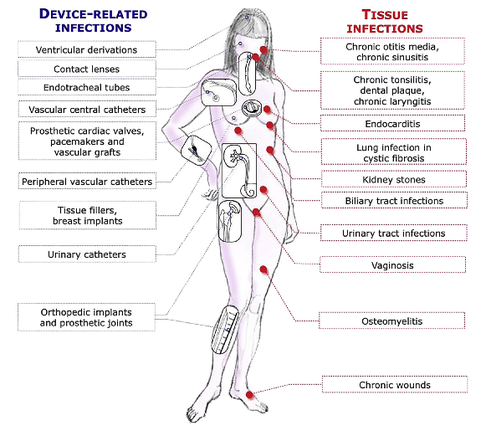
The European Society for Clinical Microbiology and Infectious Diseases has now released new guidelines on the diagnosis and treatment of biofilm infections. Written for clinical microbiologists and infectious disease specialists this paper is a MUST READ for anyone involved in treating critically ill patients.

These guidelines outline the nature and properties of biofilms and and their implications on mostly chronic infections caused. As biofilms are very common in critically ill patients it is important to know what specific problems you might encounter, how to proceed and perform a proper diagnosis and what are the essential bits and pieces in the prevention and treatment of biofilm infections.


The article is OPEN ACCESS:
Clin Microbiol Infect. 2015 Jan 14. pii: S1198-743X(14)00090-1.

Picture

New Guidelines for the Management of Atrial Fibrillation

13/3/2015

 
Picture
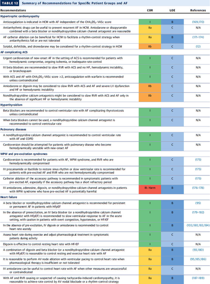
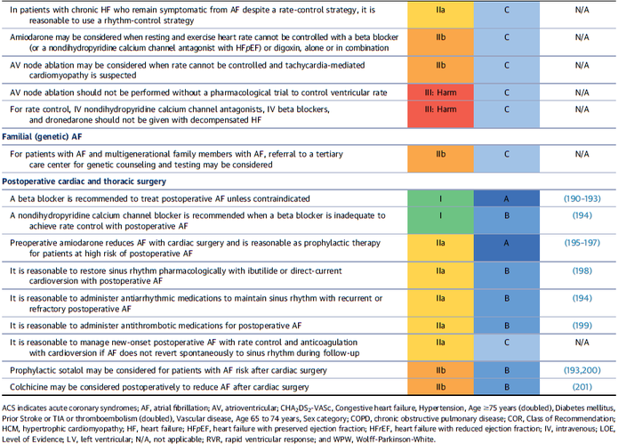
In December 2014 the AHA, ACC and HRA have released a new bunch of guidelines on the management of patients with atrial fibrillation (AFib). The paper itself is worth reading as it looks into the basic understanding of this condition, its clinical evaluation and finally the treatment options. 

As these guidelines are open access it can be considered mandatory Free Open Access Meducation FOAMed. Below is a summary of the Recommendations according to specific patient groups.

It's interesting to notice that digoxin still plays a role in patients with heart failure, especially when looking at the findings of
 Turakhia et al. in JACC, Aug 19 2014.


J Am Coll Cardiol. 2014;64(21):2246-2280    OPEN ACCESS

BIJC post on dixogin in critical care


Picture
Picture

Another Reason Why Tramadol is Not an ICU Drug

7/3/2015

 
Picture
Developed in the early 70'ies tramadol has become a very popular drug for pain relief for various reasons. Among others it is often said that tramadol is safe to use and has non-addictive properties, making this an ideal opioid to use for in and out of hospital. The facts though point in the opposite direction.

In JAMA Internal Medicine Fournier et al. have just published a case control analysis to look at the fact that tramadol before has been associated with the occurrence of significant hypoglycemia. Their cohort included a total of 334'034 patients whereas each case of hospitalization for hypoglycemia was matched with up to 10 controls on age, sex, and duration of follow-up. Basically they compared similar patients which were either started on tramadol or codeine for pain treatment. They were able to show that compared with codeine, tramadol use was associated with an increased risk of hospitalization for hypoglycemia, particularly in the first 30 days of use. It has to be noted though, that the overall incidence is low with 7 per 10'000 per annum.

In the same issue's commentary Nelson and Juurlink take the opportunity to point out some other remarkable problems associated with Tramadol, again showing us that things are not a simple as we think they are.

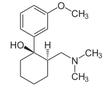
- Tramadol itself has only a low affinity to opioid receptors and mainly works over one of its metabolites: O-Desmethyltramadol (M1), which then binds to µ opioid receptors
- The expression of the enzyme that metabolites tramadol to M1 is extremely variable, thus: giving a certain dose of tramadol leaves you with an unknown dose of acting opioid!
- Despite suggestions to the contrary, tramadol does pose a risk for addiction
- And there are increasing reports of deaths involving this drug
- Other documented adverse effects are: serotonin syndrome and seizures


Conclusion: Tramadol remains a non-ideal drug in the setting of an ICU. 


Fournier et al. JAMA Intern Med. 2015;175(2):186-193.

Nelson and Juurlink JAMA Intern Med. 2015;175(2):194-195.

Subglottic Suctioning Prevents Harm in the Long-Term Intubated Patient - So Do it!

5/3/2015

 
Picture
Microbiologically confirmed ventilator-associated pneumonia (VAP) or ventilator-associated conditions (VAC, e.g. worsening oxygenation) in intubated patients remains a major concern in ICU's. VAP is defined as a hospital-acquired pneumonia which develops within 48-72 hours after endotracheal intubation.
To prevent this complication ICU's uniformly have adapted the VAP-bundle, a bunch of measures aiming to prevent ventilator-associated pneumonia. Unfortunately the evidence of the VAP-bundle is not as robust as one might think it is. Here's the  evidence of some elements of the VAP bundle:

- Elevation of the head to bed 45° (low evidence)
- Daily sedation interruptions (the impact on reducing VAP has not been shown so far)
- Daily oral chlorhexidine rinses (low evidence)


... it's most likely the combination of measures that is of benefit to the patient... hopefully! But hold on, there is another intervention that finally brings quite some evidence with it!

Active suctioning of the subglottic area, where nasal-oral secretions gather and create a rich culture medium for all sorts of micro-organisms, also aims to reduce the incidence of VAP. In contrast to the classical VAP-bundle the evidence here is strongly in favour for these devices! 

In 2005 four registrars in cardiothoracic surgery looked into this topic and summarised their efforts online on Best Evidence Topics, best bets.org. In this blog they review 13 relevant articles on the use of subglottic suctioning and conclude: subglottic suction significantly reduces the incidence of VAP in high risk patients - which means a NNT of 8 if ventilated for more than 3 days. They also mention that this measure is cost effective, despite the more expensive tubes.

In the same year Dezfulian et al. presented a systematic meta-analysis of randomized trials in the American Journal of Medicine. They ended up with 5 studies including 869 patients. They also came to the conclusion that subglottic secretion drainage is effective in preventing VAP in patients expected to be ventilated for more than 72 hours.

In 2011 Hallais et al. looked into the issue of cost-effectiveness with a cost-benefit analysis. Even when assuming the most pessimistic scenario of VAP incidence and costs the replacement of conventional ventilation with continuous subglottic suctioning would still be cost-effective.

In 2011 Muscedere et al. published an 'official' review article in Critical Care Medicine and also ended up with 13 randomised clinical trial, most of them the same 'BestBETs' had already identified 6 years before. It is therefore not surprising to see that they also found a highly significant reduction in VAP. They were also able to demonstrate a reduction in ICU length of stay and duration of mechanical ventilation, although the strength of this association was weakened by heterogeneity of study results.


We finally would like to mention the latest randomised controlled trial on this topic which was published in Critical Care Medicine this January 2015. Damas et al. randomly assigned 352 patients to either receive subglottic suctioning or not. Again sublottic suctioning significantly reduced VAP prevalence and therefore also antibiotic use.

At least we have identified one area in critical care where an impressive pile of evidence supporting the use of subglottic suctioning in long-term intubated patients is present... and even better: cost-effective analyses also come out in great favour for this measure!

Take-home message: Subglottic suctioning does prevent VAP in patients likely to be ventilated more than (48-) 72 hours and should be used in these situations.


Review BestBETs 2005

Dezfulian C et al. Am J Med. 2005 Jan;118(1):11-8

Hallais C. et al. Infect Control Hosp Epidemiol. 2011 Feb;32(2):131-5


Muscedere J et al. Crit Care Med. 2011 Vol. 39, No. 8

Damas P et al. Crit Care Med. 2015 Jan;43(1):22-30

Excellent Review on IV Fluid Resuscitation for the End of the Year 2014

11/1/2015

 
Picture
Again we have picked a review article looking at fluid resuscitation in the ICU. This article by Lira et al. in the Annals of Intensive Care looks at all the new literature available in regards of fluid therapy during resuscitation. Also review current recommendations and recent clinical evidence. This results in an excellent systematic review that leaves us with following conclusions:

- Currently no indications exist for the routine use of colloids over crystalloids

- In regards of current evidence (including the Albios trial), the cost and limited shelf time the use of albumin as a resuscitation fluid is not recommended

- The use of hydroxy-ethyl-starch (HES) during resuscitation should be avoided

- In light of the lack of evidence, and the theoretical potential for adverse effect, the suggestion is to avoid gelatine or dextran

- The use of 0.9% normal saline is associated with the development of hyperchloremic metabolic acidosis and increased risk of AKI in susceptible patients. Therefore balanced crystalloid solutions should be considered/ preferred

- Current literature supports the use of balanced crystalloid solutions (e.g. Hartmann's solution, Ringer's lactate) whenever possible


This makes things quite simple actually... but of course opinions differ!


Lira and Pinsky, Annals of Intensive Care Dec 2014, 4:38     OPEN ACCESS

Read here: The Albios trial
Little Christmas Present: Open Access 'Anaesthesia' Supplement on Transfusion, Thrombosis and Bleeding Management

27/12/2014

 
Picture
'Anaesthesia', the Journal of the Association of Anaesthetists of Great Britain and Ireland have published an Open Access Supplement on various aspects of Transfusion, Thrombosis and Bleeding Management. This is an excellent opportunity to update your knowledge in this field and actually compulsory for anyone involved actively in critical care. 

The supplement consists of multiple review articles which are kept nice and short and are perfect for reading in between...

In Conclusion: Reading highly recommended!


On following website you can find a list of all articles including links to the full text:

Anaesthesia, Vol. 70, Issue Supplement s1, January 2015: Transfusion, Thrombosis and Bleeding Management


<<Previous
Forward>>

    Search


    ​Translate Page

    Select your language above. Beware: Google Translate is often imprecise and might result in incorrect phrases!

    Picture


    ​Categories

    All
    Airway
    Cardiovascular
    Controversies
    Endocrinology
    Fluids
    For A Smile ; )
    Guidelines
    Infections
    Meducation
    Neurology
    Nutrition
    Pharmacology
    Procedures
    Radiology
    Renal
    Respiratory
    Resuscitation
    SARS CoV 2
    SARS-CoV-2
    Sedation
    Sepsis
    Transfusion

    Archives

    December 2025
    June 2025
    January 2021
    September 2020
    March 2020
    February 2020
    January 2020
    December 2019
    November 2019
    July 2019
    May 2019
    March 2019
    February 2019
    January 2019
    December 2018
    January 2018
    October 2017
    August 2017
    June 2017
    March 2017
    February 2017
    January 2017
    October 2016
    July 2016
    June 2016
    April 2016
    February 2016
    December 2015
    October 2015
    September 2015
    August 2015
    July 2015
    June 2015
    May 2015
    April 2015
    March 2015
    January 2015
    December 2014
    November 2014
    October 2014
    September 2014
    August 2014
    July 2014
    June 2014
    May 2014
    April 2014
    March 2014
    February 2014
    January 2014
    December 2013
    November 2013

    Author

    Timothy Aebi

    RSS Feed

ABOUT US

The Crit ☁​ supports free access medical education. The content of this website can be used and reproduced by stating a reference to our site.
Read our statements here

SEARCH US

CONTACT US

If you have any interesting article or news to be posted on this website, please contact us here
​

FOLLOW US

Non-AI-Website

These Blog-Posts are not written by Artificial Intelligence AI. There might be some mistypings or grammatical mistakes - but therefore no AI-writing involved!
Picture