CRIT CLOUD

  • Summaries & Reviews
    • Airway
    • Cardiovascular
    • Controversies
    • Endocrinology
    • Fluids
    • Guidelines
    • Infections
    • Neurology
    • Nutrition
    • Pharmacology
    • Procedures
    • Radiology
    • Renal
    • Resuscitiation
    • Respiratory
    • Sedation
    • Sepsis
    • Transfusion
  • World of Foam
  • FOAM.education
  • Education
    • ACLS Training ICU Lindenhofspital
    • Download Presentations
    • Download CME Galway Clinic 🔒
    • Download CME Lindenhof Hospital 🔒
    • Echo 🔒
    • Presentations🔒
    • Multimedia
  • About Us
    • Contact

Reviews and Summaries

@ILCOR 2020: Let's Put the Supraglottic Airway First!

13/2/2020

 
Picture

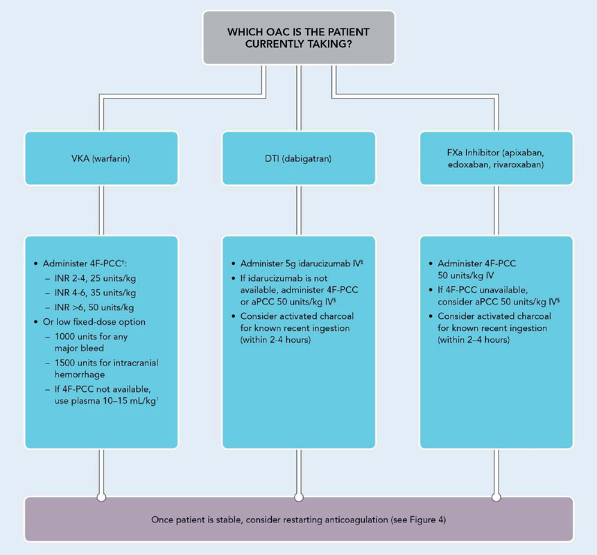
​The International Liaison Committee on Resuscitation has published the last guidelines for advanced cardiac life support (ACLS) on resuscitation ILCOR in 2015. Usually, these statements are updated every five years, but 'Circulation' has now published an AHA (American Heart Association) focused update due to an increased number of studies looking at ACLS-specific interventions.


These updates are focused on three specific areas:
  1. Advanced airway management
  2. Vasopressors
  3. Extracorporeal cardiopulmonary resuscitation ECPR


​No News in regards to Vasopressors and ECPR

Vasopressors in Cardiac Arrest


  • Epinephrine (aka Adrenaline) should be administered to patients with cardiac arrest (Class I; Level of Evidence B-R)
  • It is reasonable to administer 1mg every 3 to 5 minutes (Class IIa; Level of Evidence C-LD)
  • High-dose epinephrine is not recommended for routine use in cardiac arrest

The bottom line:​ Great, these recommendations are no real news and do not change current guidelines at all.


Extracorporeal Cardiopulmonary Resucitation ECPR

  • There is insufficient evidence to recommend the routine use of ECPR for patients with cardiac arrest AND ECPR may be considered for selected patients as rescue therapy when conventional CPR efforts are failing in settings in which it can be expeditiously implemented and supported by skilled providers

The bottom line: ECPR is not for on the roads and remains an exception in general.


Advanced Airway Management

Taking recent evidence into account the updated guidelines 2019 conclude:
​
  • Either BMV or an advanced airway strategy may be considered during CPR for adult cardiac arrest in any setting (Class 2b; Level of Evidence B-R).
  • If an advanced airway is used, the SGA can be used for adults with OHCA in settings with low tracheal intubation success rate or minimal training opportunities for ETT placement (Class 2a; Level of Evidence B-R).
  • If an advanced airway is used, either the SGA or ETT can be used for adults with OHCA in settings with high tracheal intubation success rates or optimal training opportunities for ETT placement (Class 2a; Level of Evidence B-R).
  • If an advanced airway is used in the in-hospital setting by expert providers trained in these procedures, either the SGA or ETT can be used (Class 2a; Level of Evidence B-R).
  • Frequent experience or frequent retraining is recommended for providers who perform ETI (Class 1; Level of Evidence B-NR).
  • Emergency medical services systems that perform prehospital intubation should provide a program of ongoing quality improvement to minimize complications and to track overall SGA and ETT placement success rates (Class 1; Level of Evidence C-EO).​
​

We Suggest: Put the Supraglottic Airway First!


​In regards to these updated guidelines, the necessity of optimal cardiopulmonary resuscitation (CPR) during resuscitation and practical considerations, it seems reasonable to put the supraglottic airway (SGA) to the very top of airway management!
​Here is why:


  • During resuscitation maintaining circulation and therefore vital coronary perfusion pressure (CPP) is the mainstay of success
  • BMV requires interruptions of CRP (30:2),  this is deleterious!
  • Avoiding unnecessary interruption of compressions remains therefor a top priority. Interruptions result in the sudden collapse of CPP, which will hinder successful CPR ​​
Picture
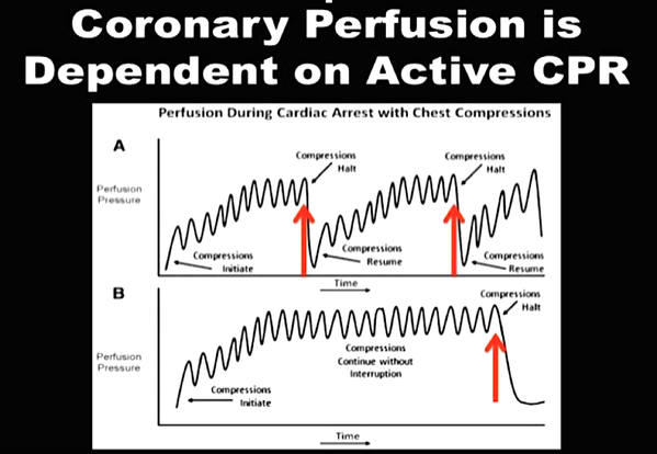
Avoiding interruptions is the key to successful CRP and therefore survival
​
  • Bag mask ventilation (BMV) can be quite tricky, especially when performed by untrained personnel. 
  • BMV is NOT a secure airway; the risk of aspiration is significant!

On the other hand

  • While providing a 'secure' airway, successful endotracheal intubation requires skilled hands and regular training
  • ETI's are mostly outside the scope of practice among many doctors, nursing staff or paramedics
  • Intubations under CPR conditions are never easy and might be even more challenging out-of-hospital
  • Again, CPR is often interrupted to provide optimal conditions for endotracheal intubation

It, therefore, seems plausible to put the supraglottic airway first. Not only first as a choice of airway management, but also one of the first things to do:

  • Placing a supraglottic airway (SGA) is simple and straight forward. Anyone can learn this procedure in a short time. We teach ICU doctors and nurses successfully on how to use non-inflatable supraglottic airways (e.g. the i-Gel device) for CPR.
  • Placing an SGA is easier than simple bag-mask ventilation (BMV)!
  • An SGA allows continuous compressions and ventilation simultaneously - no need for deleterious interruptions
  • An SGA protects the airway from aspiration fairly well - some devices even allow the introduction of a small suction catheter into the stomach
  • Moreover, if required, endotracheal intubation can still be performed by using a bougie through the SGA. This provides another option to perform ETI without interruptions of chest compressions.
  • And last but not least, SGA's allow continuous measurement of end-tidal CO2 ​
Picture
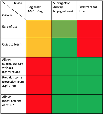
Summary of Evidence and Experience on Airway-Devices used for CRP

The International Liaison Committee on Resuscitation (ILCOR) has again carried together all evidence and recently published more than 50 new ILCOR treatment recommendations and scoping reviews. You can find these documents right here: https://costr.ilcor.org 

This website provides an excellent systematic review of the Advanced Airway Management during Adult Cardiac Arrest, containing references to all relevant evidence available.


Picture


​Based on this and given the experience from everyday clinical practice, it would be worth considering supplementing the recommendations as follows.

- For resuscitation performed by health care professionals (physicians, nurses, paramedics), the use of a supraglottic airway (ideally non-inflatable) as soon as possible is recommended.


2019 AHA Focused Updated on Adult Cardiovascular Life Support

​

Has Levosimendan Failed? - A Review of Current Evidence

11/5/2019

 
Picture

​Acute decompensated heart failure comes in many ways as remains a challenge for optimal treatment since various conditions can cause it, e.g. advanced chronic heart failure, cardiogenic- and septic-shock, cardiac- and non-cardiac surgery, etc. Among many other interventions, the infusion of positive inotropes is often used on one side and vasodilators on the other side to stabilize the situation and improve cardiac function. Despite all these measures, the re-admission rate and mortality remain a significant problem among these patients. 

While Levosimendan is still under investigation in the U.S., it is used worldwide for the short-term treatment of acutely decompensated severe chronic heart failure, especially when other treatment options have failed. Some recommend the usage of this drug in a different setting like sepsis-related heart failure or coronary heart disease.


How does Levosimendan work?


Levosimendan has some remarkable properties, which are mainly caused by three mechanisms:

1. Enhancement of the calcium sensitivity of the myofilament by binding to troponin C.

2. Opening of adenosine triphosphate-sensitive potassium (KATP) channels in vasculature smooth muscle.

3. Opening of mitochondrial KATP channels.


These mechanisms result in a positive inotropy of the heart, an increase in stroke volume (SV) and therefore, cardiac output (CO). Besides, it's vasodilatory properties seems beneficial for coronary perfusion and reduce pulmonary capillary wedge pressure (PCWP).

All these effects do not appear to induce an unfavourable energy balance in the myocardial cell, and also the oxygen demand is not increased.
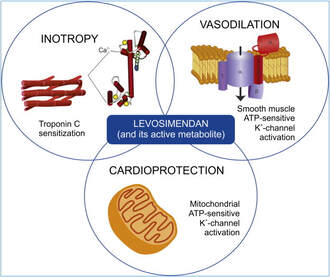
Picture
ScienceDirect.com


​So what's the Evidence?
​
The Small Bits and Pieces


​First publications back in the 90ies and early 2000s were indicative for the properties of levosimendan.  Three studies were randomized and double-blinded but very low in patient numbers and therefore not powered enough to provide substantial evidence. Their statements were also not concerning patient outcome or mortality, but its results confirmed that levosimendan enhances cardiac output without oxygen wasting, is well tolerated and leads to favorable hemodynamic effects.


Lilleberg et al. Eur Heart J. 1998;19:660–668.

Ukkonen et al. Clin Pharmacol Ther. 2000;68:522–531.

Nieminen et al. J Am Coll Cardiol. 2000;36:1903–1912.


In 2003 Kivikko et al. published further data from another small trial indication that its beneficial effects (decreases in left and right heart filling pressures and in SVR, as well as increases in stroke volume and cardiac index) are maintained for at least 24 hours after discontinuation of a 24-hour infusion. At this point, it is worth mentioning that the author published these findings as a current employee of Orion Pharma, which manufactures levosimendan.

Kivikko et al. Circulation. 2003;107:81–86.


Further data indicated that its effect might be sustained for up to at least a week, although also this study included only 22 patients!

8. Lilleberg et al. Eur J Heart Fail. 2007;9:75–82.


A Lancet publication in 2002 by Follath et al. presented a multicentre, randomized, double-blind trial in which they compared the effect of levosimendan to dobutamine in patients that were admitted to a hospital with low-output heart failure and were judged to require hemodynamic monitoring and treatment with an intravenous inotropic agent. In these 203 patients, levosimendan had a consistently better effect than dobutamine on the individual hemodynamic variables at the end of the 24 h treatment period (cardiac output and pulmonary capillary wedge pressure). The change in clinical symptoms like fatigue and dyspnoea were not significantly different, though. 
​

Interestingly, for the first time, the levosimendan group showed lower mortality than in the dobutamine group for up to 180 days.​ However, it's important to mention its absence of placebo control and its rather small sample size.

Follath et al. Lancet. 2002;360:196–202.


The Big Lumps

The SURVIVE Study


In the SURVIVE study Mebazaa et al. compared the efficacy and safety of intravenous levosimendan or dobutamine in patients hospitalized with acute decompensated heart failure who required inotropic support.
They performed a

​randomized, double-blind trial at 75 centers in 9 countries

in which they evaluated

1327 patients hospitalized with acute decompensated heart failure who required inotropic support

They found that

the addition of levosimendan to standard therapy resulted in fewer deaths compared with dobutamine, especially in the first few weeks after treatment

but

Levosimendan did not significantly reduce all-cause mortality at 180 days or affect any secondary clinical outcomes
Mebazaa et al. JAMA. 2007 May 2;297(17):1883-91.


The REVIVE Studies - Heart Failure


In the Randomized Multicenter Evaluation of Intravenous Levosimendan Efficacy studies (REVIVE I and REVIVE II) the efficacy of levosimendan on symptoms of heart failure during five days after starting a 24h trial drug infusion was assessed. 

Each patient's clinical course over 5 days was determined by a composite of the patient's self-assessment of symptoms together with a physician's assessment of the occurrence of clinical deterioration. 
​
The primary endpoint of the study was a new clinical composite endpoint (improvement, unchanged or worse - including death) derived from studies in chronic heart failure and first evaluated in 100 ADHF patients in the REVIVE I pilot study.


In REVIVE II the investigators performed one of the

first large, prospective, randomized, double-blind, controlled trials

in which they evaluated

600 patients  admitted at 103 sites in the United States, Australia, and Israel

with 

worsening heart failure and dyspnea at rest despite treatment with intravenous diuretics, and left ventricular ejection fraction of < 35% measured within the last year

whereas

patients were randomized to either receive levosimendan for 24h or standard treatment alone​

They found that after 5 days

- more patients receiving levosimendan experienced improvement compared with those who were on placebo (19.4% vs 14.6%, respectively, a 33% relative increase; P = .015)

- fewer patients receiving levosimendan worsened compared with patients who were on placebo (19.4% vs 27.2%, respectively), a 29% relative decrease, and

- fewer patients receiving levosimendan required rescue therapy (15.1%) vs placebo (26.2%), a 42% relative decrease.


Among other secondary endpoints, levosimendan improved

B-type natriuretic peptide (BNP) levels, length of hospital stay and dyspnoea at 6 hours

But

 mortality at 90 days did not differ significantly between treatment arms (a secondary endpoint) and

the most common treatment-emergent cardiovascular adverse events were more frequent with levosimendan, including hypotension (50% vs 36%), ventricular tachycardia (25% vs 17%), and atrial fibrillation (8% vs 2%).​

JACC Heart Fail. 2013 Apr;1(2):103-11. doi: 10.1016/


The LeoPARDS-Trial - Sepsis


In this study investigators wanted to know whether in adult patients who have sepsis the application of levosimendan reduces the incidence and severity of acute organ dysfunction compared with placebo.
They performed a

Randomised, double-blind, placebo-controlled multi-centre trial in 34 general ICUs in the UK

in which they evaluated

516 patients with suspected or confirmed infection and 2 or more SIRS criteria who were dependent on vasopressors for at least four hours to maintain their blood pressure.

They compared 

the intravenous infusion of levosimendan or placebo for 24 hours in addition to standard care

and found:

- No significant difference in the daily Sequential Organ Failure Assessment (SOFA) score up to day 28 (primary outcome)


- No statistical difference on death at 28 days, at ICU discharge and hospital discharge (secondary outcome)

- And: The use of levosimendan was associated with more hemodynamic instability, lower mean arterial pressures, therefore more need for noradrenalin at 24h and significantly more supraventricular tachyarrhythmias (all secondary outcomes).
N Engl J Med 2016; 375:1638-1648
​


​The CHEETAH Trial - Cardiac Surgery


The question in CHEETAH was if levosimendan compared to placebo reduces mortality in patients undergoing cardiac surgery with left ventricular dysfunktion.


This time Landoni et al. performed a

Multi-centre, randomised, placebo-controlledand parallel group designed trial 

in which they evaluated 

506 patients scheduled for cardiac surgery with peri-operative cardiovascular dysfunction 

defined as

pre-operative left ventricular ejection fraction (LVEF) < 25%, pre-operative intra-aortic balloon pump (IABP), intra- or post-operative (within 24 hours) IABP or significant inotropic requirement

They compared

Levosimendan infusion continued for up to 48 hours (or until ICU discharge) or placebo

and found

No difference in mortality at thirty 30 days (primary outcome)

And no difference in survival over time, renal replacement therapy, the median duration of mechanical ventilation, the median hospital stay and interruptions due to adverse events (all secondary outcomes).
N Engl J Med 2017; 376:2021-2031

​

Any Other Clues?


In contrast to these clinically somewhat discouraging results Shang et al. have published a meta-analysis in 2017 of randomized controlled trials looking at the usage of levosimendan in patients with heart failure, cardiogenic shock and acute coronary syndrome. 

They ended up looking at a total of nine studies, most of them low in patient numbers and comparing levosimendan to either placebo or other drugs (dobutamine and enoximone).
Picture

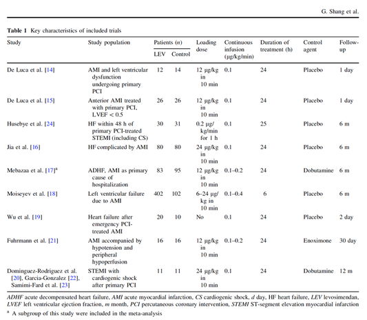
According to these data the authors conclude that levosimendan is associated with reduced total mortality, decreased incidence of worsening HF, and improved hemodynamic outcomes and does not increase the risk of adverse events except for hypotension in patients with HF (including CS) complicating ACS. Thus, levosimendan should be recommended for routine clinical application in these patients.

Am J Cardiovasc Drugs 
2017 Dec;17(6):453-463.
​
Picture


According to current evidence

- Levosimendan has interesting mechanisms of action and successfully seems to enhance cardiac output without additional oxygen wasting. This includes decreased right and left ventricular filling pressures, decreased SVR,  as well as increases in stroke volume and cardiac index. Also, PCWP is decreased.

- In terms of various patient groups, patients with acute heart failure or acute on chronic heart failure appear to benefit in terms of clinical symptoms, if at all.

- Unfortunately, there is no convincing clinical evidence that levosimendan has any benefit in long term outcomes in terms of mortality.

- And, levosimendan seems to be associated with some potential treatment-emergent cardiovascular adverse events like hypotension, supraventricular and ventricular arrhythmia.
​

Acute myocardial infarction - It's pain radiating to the right arm we have to worry about!

15/3/2019

 
Picture
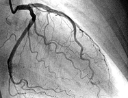
​It was only back in in 1987 when William Heberdeen (not to confound with William Heberdeen, a London doctor who described the Heberdeen nodes back in the 18th century) published the first description of ischemic chest pain. It was the birth of the classical image of strangling chest pain that occasionally radiates to the left arm, associated with exertion and relieved by rest.

A recent publication in the BMJ shows, that positive troponin levels are found frequently in patients with non-cardiac problems. This finding underlines the importance of good history taking, including the assessment of chest pain (CP) characteristics. 

​

Picture
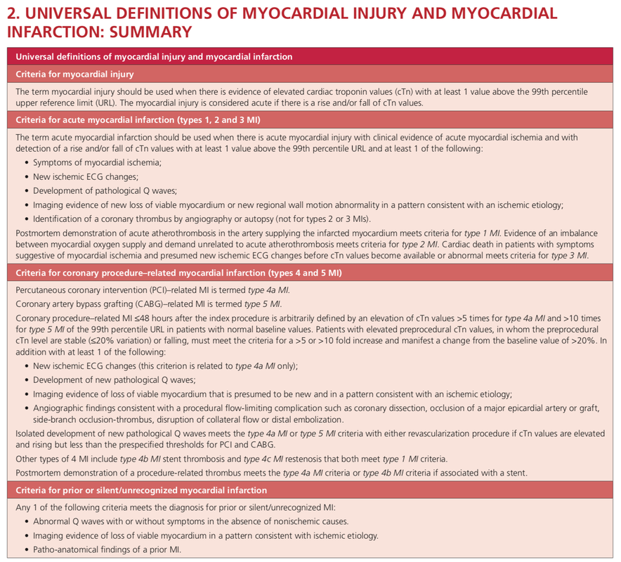
Circulation. 2018;138:e618–e651. (Click image for link to article)


​
The TRAPID-AMI Study
​

McCord J et al. have taken a closer look at chest pain and associated symptoms and their association with the diagnosis of acute myocardial infarction. They also analysed these symptoms in relation to the size of the AMI.

They performed an


international, multicenter diagnostic study

in which they evaluated 

1282 patients admitted for possible AMI to emergency departments in Europe, the US and Australia

The outcome was

the correlation of symptoms with the diagnosis of AMI and its relation to myocardial infarction size

They found that

1. only 4 symptoms were independently predictive of AMI

- Radiation to the right arm/ shoulder (OR 3.0, CI 1.8-5.0)
- Chest pressure (OR 2.5, CI 1.3-4.6)
- CP worsened by physical activity (OR 1.7, CI 1.2-2.5)
- Radiation to left arm/ shoulder (OR 1.7, CI 1.1-2.4)


2. Patients with more than 1 of the 4 symptoms were more likely to have AMI
- For patients who had all 4 symptoms, 55% had a diagnosis of AMI


3. Patients with larger AMI's were more likely to have 
- pulling CP
- pain in the right upper chest (right supramammillary area)
- and right arm/shoulder radiation


​
Picture


​- It's time to get rid of the classical image of CP radiating only into the left arm. While this might still be the predominant complaint at presentation, it's the right sided shoulder/ arm pain we should keep a close eye on!

- Chest pain radiating to the right shoulder/ arm is more predicitive of myocardial infarction than left sided chest pain

- And remember: a positive Troponin alone does not fullfill the diagnostic criteria of AMI!


Crit Pathw Cardiol. 2019 Mar;18(1):10-15.
​

Manage Critical Bleedings in Anticoagulated Patients like a Pro!

5/12/2018

 
Picture
Anticoagulated patients are common, and the amount of available oral anticoagulants is becoming more diverse and confusing. Anticoagulation is the cornerstone in the treatment of thrombosis and thromboembolic complications in a variety of diseases. Lixiana, Pradaxa, Eliquis and Xarelto are some of these pretty-sounding drugs that many doctors know but find it difficult to keep up.

So if you work in an emergency room, anaesthesia or intensive care, there's a good chance you will be facing an anticoagulant patient with potentially critical bleeding that could require urgent treatment... And this leaves you with the following questions:​

- What is a critical bleed (apart from obvious massive bleeding)? Does this bleeding need imminent reversal?

- Do I need any laboratory testing before?

- What treatment should I actually give the patient?


If you do not have a guideline in your institution, it may be time to create one, and the following publication is indeed very useful for this purpose!​

The 2017 ACC Expert Consensus Decision Pathway on Management of Bleeding in Patients on Oral Anticoagulants very nicely summarises current evidence and expert opinion on these issues. But the very best are their excellent figures, providing all the answers you need: simple and very understandable!

What is a Critical Bleeding?

Picture


​Do I Need any Laboratory Tests Before?

Picture
Picture


​What Treatment Should I Give the Patient for Reversal?

Picture


2017 ACC Expert Consensus Decision Pathway on Management of Bleeding in Patients on Oral Anticoagulants, Volume 70, Issue 24, December 2017

Out-of-Hospital Cardiac Arrest: The Power of Adrenaline and Amiodarone

29/7/2016

 
Picture
For the resuscitation out-of-hospital one of the mainstays besides compression and defibrillation ist the application of adrenalin and amiodarone. According to the new ACLS guidelines 2015 these are the only drugs remaining in the treatment for shockable rhythms.

​While adrenaline is given for maximum vasoconstriction in order to promote coronary perfusion pressure CPP, amiodarone and sometimes lidocaine are used to promote successful defibrillation of shock-refractory ventricular fibrillation VF or pulseless ventricular tachycardia VT. While the usage of these drugs is undoubtedly very effective in patients with existing circulation the effectiveness during resuscitation remains a matter of debate.

The Effect of Adrenaline

As a matter of fact it has never been proven that adrenalin actually improves long-term outcome. In 2014 Steve Lin and colleagues published a systemativ review on the efficacy of adrenaline in adult out-of-hospital cardiac arrest (OHCA). They were able to show that according to current evidence standard dose adrenaline (1mg) improved rates of survival to hospital admission and return of spontaneous circulation (ROSC) but had no benefit in means of survival to discharge or neurologic outcomes.

What about Amiodarone and Lidocaine?


Kudenchuck et al. now made the effort to look into the efficacy of amiodarone and lidocaine in the setting of OHCA. Used according to the ACLS guidelines 2016 amidarone is given after the third shock applied when treating a shockable rhythm. Two rather small controlled trials have shown so far that using amidarone actually does increase the likelihood of ROSC and the chance to arrive at a hospital alive. It's impact on survival to hospital discharge and neurologic outcome though remains uncertain.

In this randomized, double-blind trial, the investigators compared parenteral amiodarone, lidocaine and saline placebo in adult, non-traumatic, OHCA. They ended up with 3026 patients meeting inclusion criteria and which were randomly assigned to receive amiodarone, lidocaine or saline placebo for treatment. They finally found that neither amiodarone nor lidocaine improved rate of survival to discharge or neurologic outcome significantly. There were also no differences in these outcomes between amiodarone and lidocaine. Across these trial groups also in-hospital care like frequency of coronary catheterisation, therapeutic hypothermia and withdrawal of life-sustaining  treatments did not really differ, making a bias due to treatments after admission unlikely.

Take Home

- This study was not able to show any benefit of amiodarone or lidocaine in the the setting of OHCA  in terms of survival to hospital discharge and neurologic outcome

- Amiodarone seems to improve the likelihood of ROSC and survival to hospital admission (similar to adrenaline)

- As there are no other options, I believe amiodarone should remain part of the standard treatment for shockable rhythms in OHCA

- Lidocaine can be safely removed from CPR sets as there is no benefit of over amiodarone

​
Read here:

N Engl J Med 2016;374:1711-22

Resuscitation, June 2014, Vol 85, Issue 6, p 732-740


New ACLS Guidelines 2015, The Changes

Lactate - From Bad to Good? An Explanation Trial

14/2/2016

 
Picture
The discussion on the so-called lactic acidosis and its causes have become increasingly attractive over the last couple of years as several biochemical explanations are challenged. A significant confusion persists on the various relationships between lactate, lactic acid and metabolic acidosis. 

Most clinicians continue to refer to the traditional understanding of impaired tissue oxygenation causing increased lactate production, impaired lactate clearance and therefore resultant metabolic acidosis. Just recently we had a discussion on our ward round on this topic when a team member presented the most recent article of UpToDate online on the causes of lactic acidosis. The authors state that 'Lactic acidosis is the most common cause of metabolic acidosis in hospitalised patients' and that 'Lactic acidosis occurs when lactate production exceeds lactate clearance. The increase in lactate production is usually caused by impaired tissue oxygenation...'... finally suggesting that lactate is no good!

These statements support the classical understanding that:
- Hyperlactatemia is caused by tissue hypoxemia, and
- This in turn then leads to a metabolic acidosis called lactic acidosis


This biochemical understanding has persisted for decades, but there are some good reasons to vigorously challenge this traditional aspect on the 'bad' lactate. Lactate turns out to be by far more complex in its characteristics and functions, so I decided to try and make a short but comprehensive overview of this molecule.

What is lactate?

Picture
Lactate is a small organic molecule with the chemical formula CH3CH(OH)CO2H and structurally looks like on the image to the left. It is produced in the cytoplasm of human cells mainly by anaerobic glycolysis by the conversion of pyruvate to lactate by LDH. This chemical reaction results typically in a blood lactate to pyruvate ratio of about 10:1. And while lactate is produced, NAD+ also is incurred, and this actually can accept protons itself, so does not result in acidosis itself.

Lactate arises from the production of energy by consuming glycogen and glucose.

Picture


​Where does it come from?

Picture
Typically most people think of muscles first as an origin of lactate. As a matter of fact lactate originates from many other organs, including our red blood cells. Red blood cells always produce lactate as they lack the mitochondria required to regenerate NAD+ needed for glycolysis.  In general, you can say that tissues with lots of LDH are the primary producers of lactate. Around 20mmol/kg/day of lactate are produced under normal circumstances.

Lactate is not only produced in skeletal muscle.

Muscle: 25%
Skin: 25%
Brain: 20%
RBC: 20%
Intestine: 10%

What happens with it?

Lactate is not just for nothing. After its production by anaerobic glycolysis lactate is reutilised, for instance in the liver and the cortex of the kidneys. As an example: under the influence of cortisol it is used for gluconeogenesis in hepatocytes and restores glucose and glycogen. Also, it is a part of oxidative phosphorylation in the liver, kidney, muscles, the heart and the brain. Like this lactate helps conserve glucose levels in our blood.
​
​Lactate actually serves as a fuel for oxidation and glucose regeneration and therefore is a source for energy itself.
Picture
From The Lancet Endocrinology 2013


​
​How does hyperlactatemia develop?

In general, you can assume that there is a balance between lactate production and its consumption or usage. The classical understanding that tissue hypoxia leads to overproduction and underutilisation by impaired mitochondrial oxidation is correct.

The critical point though is that lactate is also produced via aerobic glycolysis as a response to stress. This happens in septic patients, asthmatic exacerbations, trauma and other critical conditions. In these situations, the trigger for lactate production is adrenergic stimulation and NOT tissue hypoxia. There are also several other reasons for hyperlactatemia other than tissue hypoxia:


Sepsis:      Adrenergic drive
Asthma:    Adrenergic drive
Trauma:    Adrenergic drive
Cardiogenic and haemorrhagic shock: Adrenergic drive
Pheochromocytoma: Adrenergic drive
Inflammation: Cytokine drive
Alkalosis, antiretroviral medication and others


Also, there is good evidence showing that organs like the lungs are an important producer of lactate during stress. And of course in all these conditions hypoxic and non-hypoxic hyperlactatemia might also co-exist.

In critically ill patients often other reasons than tissue hypoxia are responsible for hyperlactatemia (e.g. adrenergic drive).
​

Is lactate harmful?

In contrast to the classical understanding of lactate and lactic acidosis more and more evidence comes up indicating that lactate during stress actually serves as a fuel for energy production. Various tissues, e.g. the myocardium increase their lactate uptake during stress significantly. Also, our brain consumes more lactate during stress which is used for oxidation. Research has shown that lactate infusions improve cardiac output in pigs and even in patients with heart failure. 

Experimental work on isolated muscles suggests that circulating catecholamines and development of acidic conditions during exhaustive exercise may improve muscles' tolerance to elevated K+ levels. This implies that during high-intensity activity with high extracellular K+
 and adrenaline, lactate serves as a performance-enhancing chemical, rather than being the cause of muscle fatigue.

Lactate is not harmful to our organism. On the contrary, recent compelling evidence suggests that lactate might be beneficial, rather than detrimental, during high-intensity activity and to force development in working heart and skeletal muscle.
​

Why do critically ill patients with hyperlactatemia die more often then?

In critical care hyperlactatemia indeed is a marker of illness severity and a strong indicator of mortality. This is especially true for patients with sepsis. However, as described above, hyperlactatemia often doesn't indicate hypoperfusion or tissue hypoxia. Hyperlactatemia rather reflects the severity of illness by representing the degree of our body's activation to stress. A fall in lactate concentration following treatment of critically ill patients is due to an attenuation of the stress response rather than to correction of oxygen debt.

​Hyperlactatemia reflects a severe disease and the patients' response to stress. Patients die due to their illness, not because of high lactate.
​

What about Ringer's lactate?

Picture

Ringer's lactate (RL) is not harmful in patients with hyperlactatemia.

As a matter of fact RL turns out to be superior compared to normal saline in hyperlactatemia, acidotic patients and patients with hyperkalemia.
​

The bottom line

- Lactate is an indicator of stress, a marker of illness severity and a strong predictor of mortality, but not harmful as a molecule itself.

- Lactate is helpful
 as an essential source of energy and an important fuel for oxidation and glucose generation.

- During conditions like septic shock, there is no proof that lactate is produced only due to tissue hypoxia. In fact, well-ventilated lungs provide a large amount of lactate during sepsis. Lactate in sepsis and other critical conditions is mostly not due to hypoxemia or hypoperfusion.

- Ringer's lactate contains sodium lactate, but not lactic acid. Lactate itself, as mentioned above, is beneficial in severe disease. Therefore RL remains the fluid of choice during severe disease like for instance septic shock.

- Ringer's lactate is superior to normal saline in patients with metabolic acidosis, hyperlactatemia and also hyperkalemia.
​

Got interested in some better understanding? START READING HERE:

Emmettt et al. UpToDate online, August 2015, Causes of lactic acidosis

Garcia-Alvarez et al. Critical Care 2014, 18:503


Marik PE, Bellomo R. OA Critical Care 2013 Mar 01;1(1):3

Garcia-Alvarez et al. Lancet Diabetes Endocrinol. 2014 Apr;2(4):339-47.

Andersen JB et al. Journal of Experimental Biology  
2007  210: vii doi: 10.1242/jeb.001107​

Bakker J et al. Intensive Care Med (2016) 42:472–474


Also, have a listen to Bellomo's review on lactate:
Picture
Click in image to listen to podcast

OUT NOW: New and Updated ILCOR 2015 Treatment Recommendations on Cardiopulmonary Resuscitation

23/10/2015

 
Picture
Since the year 2000 the International Liaison Committee on Resuscitation (ILCOR) continues to evaluate all evidence and updates their recommendations in 5-year cycles. The most recent ILCOR 2015 International Consensus Conference was held in Dallas last February and the new treatment recommendation are out now.

Resuscitation remains one of the most challenging situations in health care. Providing basic and advanced cardiac life support gives you the opportunity to virtually safe a patients life but in a very limited period of time. It is an enormous challenge to consider all emerging evidence and pack this into simple and useful guidelines.

It is imperative to for any health care provider to get familiar with the updated guidelines and major changes. Below you can find all relevant links to get the reading going. 

The team of BoringEM.org in Canada have provided some excellent infographics to visualise all important changes in the new treatment guidelines since 2010. You should also note that the Canadian Heart & Stroke Association and the American Heart Association have just published the 'HIGHLIGHTS of the 2015 American Heart Association Guidelines Update for CPR and ECC', an excellent summary of the new recommendations and changes. So if you can't find the time to read all of the publication in 'Circulation', this will certainly provide all information you need to know.


Summary of the Canadian Heart & Stroke Association and the American Heart Association: HIGHLIGHTS of the 2015 American Heart Association Guidelines Update for CPR and ECC

​
​
The original publication in Circulation, October 20, 2015, Volume 132, Issue 16 suppl 1

OPEN ACCESS


The Most Important Changes (Click to Enlarge)


The Updated Algorithms (Click to Enlarge)
​


​ERC and ESICM 2015 Guidelines for Post-Resuscitation Care
​

​Based on the the 2015 ILCOR treatment recommendations the European Resuscitation Council (ERC) and the European Society of Intensive Care Medicine (ESICM) have produced these post-resuscitation care guidelines on October the 13th. Recent changes here are the greater emphasis for urgent PCI when indicated, target temperature management at 36°C, prognostic evaluation using a multimodal strategy and an increased emphasis on rehabilitation after survival.
​
​
Nolan JP, Resuscitation, October 2015, Pages 202 - 222

​

Don't Throw Away your Cooling Devices!

20/8/2015

 
Picture
When performing a kidney transplantation nowadays up to 50% of recipients developed a delayed graft function which is defined as the need of dialysis within seven days. The authors of this recently published NEJM-article asked themselves whether mild hypothermia might influence outcome in this regard. 

In order to answer this question the investigators assigned organ donors after declaration of death according to neurologic criteria into two groups. They were either treated with mild hypothermia (34 to 35°C) or with normothermia (36.5 to 37.5°C). The target temperature was maintained until the patients were transferred to theatre for transplantation. 

Primary outcome of this trial was delayed graft function among recipients. Secondary outcomes  included the rates of individual organs transplanted into each treatment group at the total number of organs transplanted from each donor. 

This trial had to be stopped early as an interim analysis showed significant efficacy of mild hypothermia. Up to this point a total of 572 patients received a kidney transplant (285 in the hypothermia group and 287 in the normothermia group). 28% of recipients in the hypothemia group developed delayed graft function compared to 39% in the normothermia group.

The authors therefore conclude that mild hypothermia significantly reduces the rate of delayed graft functions among recipients. 

  • This study suggests that potential organ donors after declaration of death according to neurologic criteria should be treated with mild hypothermia. 

  • Intensive care units that continue to treat patients with mild hypothermia after cardiac arrest might have two rewarm their patients for the diagnosis of neurological death before re-cooling them for organ transplantation

Anyhow, it seems reasonable not to get rid of your cooling devices!


Picture
Read more about the controversies of hypothermia in the ICU:


The Targeted Temperature Management Trial: Nielsen N, et al. New Engl J Med. 2013 Dec;369(23):2197-206

The 2 trials that introduced therapeutic hypothermia into ICU practice:
The Hypothermia After Cardiac Arrest Study Group, Holzer at al. New Engl J Med. 2002 Feb;346(8):549-556

Bernard S.A. et al. New Engl J Med. 2002 Feb;346(8):557-563

Review article on therapeutic hypothermia for non-VF/VT cardiac arrest:
Sandroni S. et al. Crit Care Med; 2013;17:215

Pyrexia and neurological outcome:
Leary M. et al. Resuscitation. 2013 Aug;84(8):1056-61


BIJC post on: The Effect of Pre-Hospital Cooling: Rather Worrying Results


Another Hole in the Ballon (-Pump)!

7/5/2015

 
Picture
In the late 1960's the technology of counter-pulsation by using an intra-aortic balloon pump (IABP) was introduced into clinical work. Based on the principle of diastolic inflation and systolic deflation, IABP counter-pulsation improves diastolic coronary artery blood flow and decreases left ventricular afterload. Up to the year 2009, 2012 respectively, the usage of an IABP in patients with ST-segment elevation myocardial infarction and cardiogenic shock was considered a class IC recommendation (reminder: levels of evidence).

Since then a couple of well conducted, larger trials have failed to show a positive impact of IABP especially on mortality. In regards of the most recent meta-analysis in JAMA we provide a short overview of the most important publications. It's interesting to see that the balloons undermining started with a meta-analysis and for the the time being ends with one.


Stitch no.1

The first notable hole in the ballon was caused by Sjauw et al.'s systematic review and meta-analysis in the European Heart Journal in 2009. Their pooled randomized data consisting of two separate meta-analyses did not support the use of an IABP in patients with high risk STEMI. They concluded that
there is insufficient evidence endorsing the current guideline recommendation for the use of IABP therapy in the setting of STEMI complicated by cardiogenic shock.

This publication was one of the main reasons for the expert panel of the European Society of Cardiology to change the recommendation (ESC Guidelines 2012) to use an IABP in patients with STEMI from IC to IIB.


Stitch 2 and 3

In the same year 2012 Thiele et al. published their first IABP-SHOCK II results in the NEJM. Their

randomized, prospective, open-label, multicenter trial showed no reduction in the 30-day mortality
compared to the best available medical therapy alone in patients with myocardial infarction-induced cardiogenic shock and planned early revascularization (PCI or CABG).

One year later the IABP-SHOCK II investigators published their final 12-months results in The Lancet. They came to the final conclusion that in patients undergoing early revascularization for myocardial infarction with cardiogenic shock, IABP did not reduce 12-month all-cause mortality.


Stitch no. 4

In 2013 Ranucci at al. presented the results of their
single-center prospective randomized controlled trial looking at the usage of a preoperative IABP in high-risk patients undergoing surgical coronary revascularization. By looking at a total of 110 patients with an ejection fraction below 35% and no hemodynamic instability there was no improvement in outcome when inserting an IABP preoperatively.

Preliminary Final Stitch

So finally Ahmad and his team decided to assess IABP efficacy in acute myocardial infarction by performing an updated meta-analysis. Main outcome was 30-day mortality. They included
12 eligible RCTs randomizing 2123 patients and found no improvement in mortality among patients with acute myocardial infarction... regardless of whether patients had cardiogenic shock or not!
A look at another 15 eligible observational studies with a total of 15 530 patients showed basically conflicting results which was explained by the differences between studies in the balance of risk factors between IABP and non-IABP groups.


It seems that the IABP fails to show its assumed efficacy in patients with myocardial infarction and cardiogenic shock, especially when early revascularization (PCI or CABG) is available.

As a general consideration and also when no early revascularisation is available the use of another left-ventricular assist device like the Impella pump might prove to be a good and easy to use alternative (see blow).


Sjauw KD et al. Eur Heart J 30: 459-468

ESC Guidelines for the management of acute myocardial infarction in patients presenting with ST-segment elevation. Eur Heart J 33: 2569-2619        
OPEN ACCESS

Thiele et al. N Engl J Med 2012; 367:1287-1296            OPEN ACCESS

Thiele et al. The Lancet, Volume 382, Issue 9905, Pages 1638 – 1645

Crit Care Med. 2013 Nov;41(11):2476-83

JAMA Intern Med. Published online March 30, 2015


Short film on the principle of the Impella pump 2.5. Bare in mind that this device can actually be easily inserted in the environment of ICU and positioned by using transthoracic echo TTE.


Excellent Review on IV Fluid Resuscitation for the End of the Year 2014

11/1/2015

 
Picture
Again we have picked a review article looking at fluid resuscitation in the ICU. This article by Lira et al. in the Annals of Intensive Care looks at all the new literature available in regards of fluid therapy during resuscitation. Also review current recommendations and recent clinical evidence. This results in an excellent systematic review that leaves us with following conclusions:

- Currently no indications exist for the routine use of colloids over crystalloids

- In regards of current evidence (including the Albios trial), the cost and limited shelf time the use of albumin as a resuscitation fluid is not recommended

- The use of hydroxy-ethyl-starch (HES) during resuscitation should be avoided

- In light of the lack of evidence, and the theoretical potential for adverse effect, the suggestion is to avoid gelatine or dextran

- The use of 0.9% normal saline is associated with the development of hyperchloremic metabolic acidosis and increased risk of AKI in susceptible patients. Therefore balanced crystalloid solutions should be considered/ preferred

- Current literature supports the use of balanced crystalloid solutions (e.g. Hartmann's solution, Ringer's lactate) whenever possible


This makes things quite simple actually... but of course opinions differ!


Lira and Pinsky, Annals of Intensive Care Dec 2014, 4:38     OPEN ACCESS

Read here: The Albios trial

Early Systematic Coronary Angiography in All Out-Of-Hospital Cardiac Arrests Seems Reasonable

9/6/2014

 
Picture
The most recent guidelines of the European Resuscitation Council and the American Heart Association recommend that resuscitated patients of presumed cardiac cause should undergo immediate coronary angiography with subsequent PCI if indicated, regardless of clinical symptoms and/or ECG criteria. However, this approach is discussed controversially as we don't know if there is any benefit on performing an angiography in every out-of-hospital cardiac arrest (OHCA). Additionally such an approach might be associated with quite some logistical and organisational problems for certain institutions.

In this noteworthy and open access review article Geri et al. discuss current literature and state that there are no randomised studies looking at acute coronary angiography in OHCA patients. A large number of observational studies though supports feasibility and a possible survival benefit of an early invasive approach. 

They conclude that even in the absence of large randomised studies, it is probable that an early coronary revascularization is associated with a significant clinical benefit in OHCA survivors. 

Providing the patient had no other obvious reason for OHCA (sepsis, haemorrhage, metabolic disorder etc.) current literature strongly encourages performing a systematic coronary angiography in all OHCA patients.


Geri G et al., Current Opinion in Critical Care, June 2014, Vol 20, Issue 3 (Open Access)

Challenging another Dogma: Time for Drug-Free Cardiopulmonary Resuscitation?

30/4/2014

 
Picture
Guidelines on advanced cardiopulmonary resuscitation around the world, including the ACLS guidelines, are based on four important concepts: mechanical resuscitation (CPR), defibrillation, airway management and also application of vasoactive drugs. The application of these drugs is intended to improve hemodynamics and the heart's responsiveness to defibrillation.

What some of us might not be aware of though, but has already been mentioned by the '2010 International Consensus on Cardiopulmonary Resuscitation and Emergency Cardiovascular Care Science with Treatment Recommendations' and the 'European Resuscitation Council Guidelines' in 2010: There is actually no definitive evidence that the application of these drugs provides any long-term benefit for these patients.


While vasopressors are intended to improve coronary and cerebral perfusion and therefore successful defibrillation and neurological outcome, there is actually some concern (animal and registry studies) that this might actually decrease microcirculation and cerebral blood flow as well as increase myocardial oxygen consumption and cause post-defibrillation ventricular arrhythmias. Also the use of anti-arrhythmic drugs as well as their combination with vasopressors lacks evidence of any impact on survival. Another interesting fact is that we have no idea about ideal dosage or optimal timing of these drugs... should they be given continuously?

This and many more questions and facts are discussed in an interesting review article by Sunde and Olasveengen in Current Opinion in Critical Care. They conclude that there is no evidence to support any specific drugs during cardiac arrest and that healthcare systems should not prioritise implementation of unproven drugs before good quality of care can be documented.


Are we heading towards an era of drug-free resuscitation?


Sunde K et al. Curr Opin Crit Care. 2014 April 16

Article accessible here

The Effect of Pre-Hospital Cooling: Rather Worrying Results

24/3/2014

 
Picture
JAMA just published another interesting puzzle piece on the topic of post arrest cooling. In this interesting, randomized trial, the authors addressed the question wether prehospital cooling aiming for 34°C improves outcomes after resuscitation from cardiac arrest in patients with ventricular fibrillation (VF) and non-VF. A total of 1359 patients were enrolled within 5 years. Primary outcomes were survival at hospital discharge and neurological status at discharge.

In regards of primary outcomes no differences were found between the intervention and control group. Also when looking at patients who awakened from coma or died without awakening again no significant differences between the intervention and control group were found. Also length of stay, access to early coronary angiography and reduction in level or withdrawal of life support did not differ.

When looking at patient safety issues though the investigators noted a higher incidence of rearrest during transport to the hospital. The intervention group (cooled out of hospital) also showed significantly lower oxygenation on arrival, increased pulmonary edema on the first chest x-ray and a greater use of diuretics during the first 12 hours after admission. During further hospitalization though the number of days ventilated, the incidence of re-intubation and the use of antibiotics did not differ.

The study results don’t support the practice of out-of-hospital cooling but show some worrying implications for patients safety.


Cobb LA et al. JAMA. 2013;311(1):45-52 

Open access on JAMA Network Reader (Need for sign up)

Recent BIJC blog on Targeted Temperature Management Trial: 
Targeted Temperature Management Trial: Is it Time to Stop Cooling Patients after Cardiac Arrest?

Is This the New Thrombolysis for Pulmonary Embolism?

21/2/2014

 
Picture
According to the American College of Chest Physicians (ACCP) guidelines thrombolytic therapy is not recommended for most patients (grade 1B). One reason is that controlled clinical trials have not demonstrated benefits in term of reduced mortality rates or earlier resolution of symptoms when currently compared with heparin. Another reason is that thrombolysis with tPA carries a substantial risk for severe complications like intracerebral haemorrhage. Currently accepted indications for thrombolytic therapy include hemodynamic instability or right ventricular dysfunction demonstrated on echo.

Although not scientifically proven, quite a few cardiologists will argue that thrombolysis will benefit in better clinical long term outcome by experience. The question remains whether thrombolysis is underutilized and might improve outcomes in moderate PE’s as well.

In this publication in Clinical Cardiology Mohsen at al. present an interesting concept of applying a ‘safe dose thrombolysis’ (SDT) where half the dose of tPA is given in conjunction with a modified dose of parenteral anticoagulation... rivaroxaban in this case. Over 12 months 98 patients with moderate to severe PE were treated with this regimen. They come to the conclusion that safe dose thrombolysis plus rivaroxaban was highly safe (no bleeds, recurrent VTE in 3 patients) and effective, leading to favorable early and intermediate-term outcomes and early discharge.

Despite some limitations in this publication the idea is interesting indeed and might be a first step towards a new approach towards thrombolysis.


Sharifi M et al. Clin Cardiol. 2014 Feb;37(2):78-82


Targeted Temperature Management Trial: Is it Time to Stop Cooling Patients after Cardiac Arrest?

6/1/2014

 
Picture
In 2002 two published articles in the New England Journal of Medicine changed ICU management of out of hospital arrests profoundly. According to these two articles (cited below) the American Heart Association labeled this to be good evidence (Level1) to recommend induced hypothermia in comatose survivors of out of hospital cardia arrest caused by VF. The target temperature was recommended to be between 32-34°C and to be maintained for 12-24 hours.

And now this... Nielsen et al. present the Targeted Temperature Management Trial showing, that there is NO difference between patients cooled to 33°C and patients kept at 36°C. Is this the end of the cooling era, should we change our management? 

I personally think think that this trial basically adds up to our knowledge in the field of post cardiac arrest care, but not necessarily contradicts the previous two trials. We now have one trial showing that there seems to be no difference between 33°C and 36°C but we also know, that hyperthermia (pyrexia) is troublesome and associated with worse neurological outcome. 

So, as pronounced hypothermia (33°C) makes no difference to ‘mild’ hypothermia (36°C) and pyrexia is proven to be harmful... the question is: What is the right temperature? We seem to head towards normothermia or mild hypothermia in order to provide best management for our patients. It’s going to be interesting to see how recommendations will change in the near future.


The Targeted Temperature Management Trial: Nielsen N, et al. New Engl J Med. 2013 Dec;369(23):2197-206

The 2 trials that introduced therapeutic hypothermia into ICU practice:

The Hypothermia After Cardiac Arrest Study Group, Holzer at al. New Engl J Med. 2002 Feb;346(8):549-556

Bernard S.A. et al. New Engl J Med. 2002 Feb;346(8):557-563

Review article on therapeutic hypothermia for non-VF/VT cardiac arrest:

Sandroni S. et al. Crit Care Med; 2013;17:215

Pyrexia and neurological outcome:

Leary M. et al. Resuscitation. 2013 Aug;84(8):1056-61


<<Previous

    Search


    ​Translate

    Select your language above. Beware: Google Translate is often imprecise and might result in incorrect phrases!

    Picture


    ​Categories

    All
    Airway
    Cardiovascular
    Controversies
    Endocrinology
    Fluids
    For A Smile ; )
    Guidelines
    Infections
    Meducation
    Neurology
    Nutrition
    Pharmacology
    Procedures
    Radiology
    Renal
    Respiratory
    Resuscitation
    SARS CoV 2
    SARS-CoV-2
    Sedation
    Sepsis
    Transfusion

    Archives

    January 2021
    September 2020
    March 2020
    February 2020
    January 2020
    December 2019
    November 2019
    July 2019
    May 2019
    March 2019
    February 2019
    January 2019
    December 2018
    January 2018
    October 2017
    August 2017
    June 2017
    March 2017
    February 2017
    January 2017
    October 2016
    July 2016
    June 2016
    April 2016
    February 2016
    December 2015
    October 2015
    September 2015
    August 2015
    July 2015
    June 2015
    May 2015
    April 2015
    March 2015
    January 2015
    December 2014
    November 2014
    October 2014
    September 2014
    August 2014
    July 2014
    June 2014
    May 2014
    April 2014
    March 2014
    February 2014
    January 2014
    December 2013
    November 2013

    Author

    Timothy Aebi

    RSS Feed

ABOUT US

The Crit ☁​ supports free access medical education. The content of this website can be used and reproduced by stating a reference to our site.
Read our statements here

SEARCH US

CONTACT US

If you have any interesting article or news to be posted on this website, please contact us here
​

FOLLOW US

TRUST US

TWEET US

This website is certified by Health On the Net Foundation. Click to verify. This site complies with the HONcode standard for trustworthy health information:
verify here.

Picture
Follow @BIJCorg

DISCOVER THE WORLD OF #FOAMed HERE
Picture